Radiography
[2] There are conflicting accounts of his discovery because Röntgen had his lab notes burned after his death, but this is a likely reconstruction by his biographers:[3][4] Röntgen was investigating cathode rays using a fluorescent screen painted with barium platinocyanide and a Crookes tube which he had wrapped in black cardboard to shield its fluorescent glow.
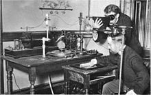
"[5] The first use of X-rays under clinical conditions was by John Hall-Edwards in Birmingham, England, on 11 January 1896, when he radiographed a needle stuck in the hand of an associate.
This was a result of Pulyui's inclusion of an oblique "target" of mica, used for holding samples of fluorescent material, within the tube.
On 3 February 1896 Gilman Frost, professor of medicine at the college, and his brother Edwin Frost, professor of physics, exposed the wrist of Eddie McCarthy, whom Gilman had treated some weeks earlier for a fracture, to the X-rays and collected the resulting image of the broken bone on gelatin photographic plates obtained from Howard Langill, a local photographer also interested in Röntgen's work.
[7] X-rays were put to diagnostic use very early; for example, Alan Archibald Campbell-Swinton opened a radiographic laboratory in the United Kingdom in 1896, before the dangers of ionizing radiation were discovered.
Initially, many kinds of staff conducted radiography in hospitals, including physicists, photographers, physicians, nurses, and engineers.
Radiographers now perform fluoroscopy, computed tomography, mammography, ultrasound, nuclear medicine and magnetic resonance imaging as well.
Since the body is made up of various substances with differing densities, ionising and non-ionising radiation can be used to reveal the internal structure of the body on an image receptor by highlighting these differences using attenuation, or in the case of ionising radiation, the absorption of X-ray photons by the denser substances (like calcium-rich bones).
The difference between soft and hard body parts stems mostly from the fact that carbon has a very low X-ray cross section compared to calcium.
[9] CT exams are generally short, most lasting only as long as a breath-hold, Contrast agents are also often used, depending on the tissues needing to be seen.
Usually the hip (head of the femur), lower back (lumbar spine), or heel (calcaneum) are imaged, and the bone density (amount of calcium) is determined and given a number (a T-score).
Fluoroscopy is mainly performed to view movement (of tissue or a contrast agent), or to guide a medical intervention, such as angioplasty, pacemaker insertion, or joint repair/replacement.
The ability to work in two planes is important for orthopedic and spinal surgery and can reduce operating times by eliminating re-positioning.
Angiography is used to find aneurysms, leaks, blockages (thromboses), new vessel growth, and placement of catheters and stents.
Industrial radiography is a method of non-destructive testing where many types of manufactured components can be examined to verify the internal structure and integrity of the specimen.
X and gamma rays have the shortest wavelength and this property leads to the ability to penetrate, travel through, and exit various materials such as carbon steel and other metals.
[16] Lead is the most common shield against X-rays because of its high density (11,340 kg/m3), stopping power, ease of installation and low cost.
[18] This initiative has been endorsed and applied by a growing list of various professional medical organizations around the world and has received support and assistance from companies that manufacture equipment used in radiology.
[19] The World Health Organization and International Atomic Energy Agency (IAEA) of the United Nations have also been working in this area and have ongoing projects designed to broaden best practices and lower patient radiation dose.
[20][21][22] Contrary to advice that emphasises only conducting radiographs when in the patient's interest, recent evidence suggests that they are used more frequently when dentists are paid under fee-for-service.
The resultant images from the radiograph (X-ray generator/machine) or CT scanner are correctly referred to as "radiograms"/"roentgenograms" and "tomograms" respectively.
A number of other sources of X-ray photons are possible, and may be used in industrial radiography or research; these include betatrons, linear accelerators (linacs), and synchrotrons.
[24] Detectors can be divided into two major categories: imaging detectors (such as photographic plates and X-ray film (photographic film), now mostly replaced by various digitizing devices like image plates or flat panel detectors) and dose measurement devices (such as ionization chambers, Geiger counters, and dosimeters used to measure the local radiation exposure, dose, and/or dose rate, for example, for verifying that radiation protection equipment and procedures are effective on an ongoing basis).
This device is made of a vacuum tube with a wide input surface coated on the inside with caesium iodide (CsI).