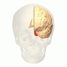
Colour centre
V1 sends the visual information received from the LGN to other extrastriate cortex areas for higher order processing.
fMRI experimental results showed that V1 has two kinds of colour sensitive neurons: single-opponent and double-opponent cells.
Double-opponent cells are particularly important in computing local cone ratios from visual information from their receptive fields.
The three types of cone cells, small (S), medium (M), and long (L), detect different wavelengths across the visible spectrum.
Recent findings have shown that the colour centre is neither isolated nor traceable to a single area in the visual cortex.
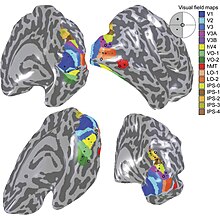
Anatomical and physiological studies have established that the colour centre begins in V1 and sends signals to extrastriate areas V2 and V4 for further processing.
[5] After identification of V4 as the colour-selective region in macaque monkeys, scientists began searching for a homologous structure in the human cortex.
A recent study from Winawer et al. analysing fMRI measurements to map the hV4 and ventral occipital areas showed variances between subjects used for hV4 mapping was at first attributed to instrumentation error, but Winawer argued that the sinuses in the brain interfered with fMRI measurements.
It was concluded that the two sub-divisions co-operate with each other in order to generate colour images, but they are also functionally separate.
[4] A study by Nunn et al. on the activation of the V4-complex in people with visual synaesthesia from hearing spoken words was used to predict the location of the colour centre.
Synaesthesia is the phenomenon where a sensory stimulus produces an automatic and involuntary reaction in a different sensation.
fMRI results showed that the left fusiform gyrus, an area consistent with V4, was activated when the subjects spoke.
[11] V2, also called the prestriate cortex, is believed to have a small role in colour processing by projecting signals from V1 to the V4-complex.
[8] V4 also has feedback on V2, suggesting that there is a defined network of communication between the multiple areas of the visual cortex.
[12] Functional magnetic resonance imaging, or fMRI for short, has been key in determining the colour selective regions in the visual cortex.
[13] Sakai et al. used fMRI to observe whether activation of the fusiform gyrus correlated with the perception of colour and the after image.
A series of three images were shown to subjects while fMRI was used to focus on the haemodynamics of the fusiform gyrus.
The results of the experiment showed that there was a significant increase of activity in the fusiform gyrus when the subject viewed the colour image.
[13] Cerebral achromatopsia is a chronic condition where a person is unable to see colour, but they are still able to recognize shape and form.
Many studies have shown that lesions in the areas commonly identified as the colour centre, such as V1, V2, and the V4-complex lead to achromatopsia.
One of the primary initiatives to locating the colour centre in the visual cortex is to discover the cause and a possible treatment of cerebral achromatopsia.
[13] The variance in symptoms emphasizes the need to understand the architecture of the colour centre in order to better diagnose and possible treat cerebral achromatopsia.