Cre recombinase
Two separate DNA species both containing loxP sites can undergo fusion as the result of Cre mediated recombination.
[2] The enzyme plays important roles in the life cycle of the P1 bacteriophage, such as cyclization of the linear genome and resolution of dimeric chromosomes that form after DNA replication.
The enzyme's ability to operate efficiently in a wide range of cellular environments (including mammals, plants, bacteria, and yeast) enables the Cre-Lox recombination system to be used in a vast number of organisms, making it a particularly useful tool in scientific research.
[4] Studies carried out in 1981 by Sternberg and Hamilton demonstrated that the bacteriophage 'P1' had a unique site specific recombination system.
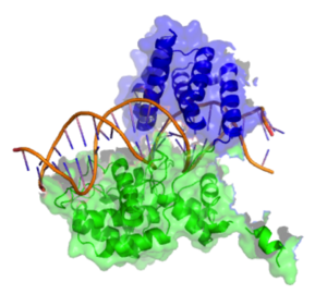
[2] Early studies also demonstrated that Cre binds to non specific DNA sequences whilst having a 20 fold higher affinity for loxP sequences and results of early DNA footprinting studies also suggested that Cre molecules bind loxP sites as dimers.
Helices A & E are involved in the formation of the recombinase tetramer with the C terminus region of helix E known to form contacts with the C terminal domain of adjacent subunits.
Crystal structures demonstrate that this terminal N helix buries its hydrophobic surface into an acceptor pocket of an adjacent Cre subunit.
If the 5’ hydroxyl groups attack the 3’-phosphotyrosine linkage one pair of the DNA strands will exchange to form a Holliday junction intermediate.
In addition to this Cre is also used to resolve dimeric lysogenic P1 DNA that forms during the cell division of the phage.
Once tamoxifen is introduced, it is metabolized into 4-hydroxytamoxifen, which then binds to the ER and results in the translocation of the CreER into the nucleus, where it is then able to cleave the lox sites.
In recent years, Cre recombinase has been improved with conversion to preferred mammalian codons, the removal of reported cryptic splice sites, an altered stop codon, and reduced CpG content to reduce the risk of epigenetic silencing in mammals.