Golgi's method
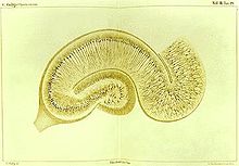
The method was discovered by Camillo Golgi, an Italian physician and scientist, who published the first picture made with the technique in 1873.
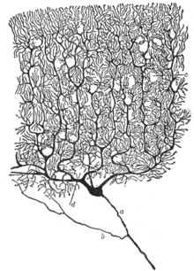
Golgi staining was used by Spanish neuroanatomist Santiago Ramón y Cajal (1852–1934) to discover a number of novel facts about the organization of the nervous system, inspiring the birth of the neuron doctrine.
Furthermore, the thin filamentary extensions of neural cells, including the axon and the dendrites of neurons, are too slender and transparent to be seen with normal staining techniques.
[2] Dendrites, as well as the cell soma, are clearly stained in brown and black and can be followed in their entire length, which allowed neuroanatomists to track connections between neurons and to make visible the complex networking structure of many parts of the brain and spinal cord.
Golgi's staining is achieved by impregnating aldehyde-fixed nervous tissue with potassium dichromate and silver nitrate.