History of neuroscience
In ancient Egypt, from the late Middle Kingdom onwards, in preparation for mummification, the brain was regularly removed, for it was the heart that was assumed to be the seat of intelligence.
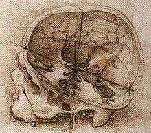
The hieroglyph for brain, occurring eight times in this papyrus, describes the symptoms, diagnosis, and prognosis of two patients, wounded in the head, who had compound fractures of the skull.
The assessments of the author (a battlefield surgeon) of the papyrus allude to ancient Egyptians having a vague recognition of the effects of head trauma.
Observations by ancient civilizations of the human brain suggest only a relative understanding of the basic mechanics and the importance of cranial security.
[3] On the opposite end, during the Hellenistic period, Herophilus and Erasistratus of Alexandria engaged in studies that involved dissecting human bodies, providing evidence for the primacy of the brain.
[2] During the Roman Empire, the Greek physician and philosopher Galen dissected the brains of oxen, Barbary apes, swine, and other non-human mammals.
He also explained phenomena such as, insomnia, mania, hallucinations, nightmares, dementia, epilepsy, stroke, paralysis, vertigo, melancholia and tremors.
He also described a condition similar to schizophrenia, which he called Junun Mufrit, characterized by agitation, behavioral and sleep disturbances, giving inappropriate answers to questions, and occasional inability to speak.
[9] Abulcasis, Averroes, Avenzoar, and Maimonides, active in the Medieval Muslim world, also described a number of medical problems related to the brain.
Between the 13th and 14th centuries, the first anatomy textbooks in Europe, which included a description of the brain, were written by Mondino de Luzzi and Guido da Vigevano.
[12] In addition to recording many anatomical features such as the putamen and corpus callosum, Vesalius proposed that the brain was made up of seven pairs of 'brain nerves', each with a specialized function.
He suggested that the pineal gland was where the mind interacted with the body after recording the brain mechanisms responsible for circulating cerebrospinal fluid.
[15] In the 1820s, Jean Pierre Flourens pioneered the experimental method of carrying out localized lesions of the brain in animals describing their effects on motricity, sensibility and behavior.
[18] In 1848, John Martyn Harlow described that Phineas Gage had his frontal lobe pierced by an iron tamping rod in a blasting accident.
[20] Broca's hypothesis was supported by Gustav Fritsch and Eduard Hitzig who discovered in 1870 that electrical stimulation of motor cortex caused involuntary muscular contractions of specific parts of a dog's body and by observations of epileptic patients conducted by John Hughlings Jackson, who correctly deduced in the 1870s the organization of the motor cortex by watching the progression of seizures through the body.
[22] Modern research still uses the Korbinian Brodmann's cytoarchitectonic (referring to study of cell structure) anatomical definitions from this era in continuing to show that distinct areas of the cortex are activated in the execution of specific tasks.
[20] Studies of the brain became more sophisticated after the invention of the microscope and the development of a staining procedure by Camillo Golgi during the late 1890s that used a silver chromate salt to reveal the intricate structures of single neurons.
Golgi and Ramón y Cajal shared the Nobel Prize in Physiology or Medicine in 1906 for their extensive observations, descriptions and categorizations of neurons throughout the brain.
In 1907, Louis Lapicque suggested that the action potential was generated as a threshold was crossed,[39] what would be later shown as a product of the dynamical systems of ionic conductances.
A great deal of study on sensory organs and the function of nerve cells was conducted by British physiologist Keith Lucas and his protege Edgar Adrian.
[41] Concurrently, Josepht Erlanger and Herbert Gasser were able to modify an oscilloscope to run at low voltages and were able to observe that action potentials occurred in two phases—a spike followed by an after-spike.
With this research, the pair discovered that the velocity of action potentials was directly proportional to the diameter of the nerve fiber and received a Nobel Prize for their work.
[42] In the process of treating epilepsy, Wilder Penfield produced maps of the location of various functions (motor, sensory, memory, vision) in the brain.
[46] Kenneth Cole joined Columbia University in 1937 and remained there until 1946 where he made pioneering advances modelling the electrical properties of nervous tissue.
[48][49] Alan Lloyd Hodgkin spent a year (1937–38) at the Rockefeller Institute, during which he joined Cole to measure the D.C. resistance of the membrane of the squid giant axon in the resting state.
Beginning in 1966, Eric Kandel and collaborators examined biochemical changes in neurons associated with learning and memory storage in Aplysia.
Eric Kandel and collaborators have cited David Rioch, Francis O. Schmitt, and Stephen Kuffler as having played critical roles in establishing the field.
[52] As a result of the increasing interest about the nervous system, several prominent neuroscience institutes and organizations have been formed to provide a forum to all neuroscientists.