History of tracheal intubation
By the late 19th century, advances in the sciences of anatomy and physiology, as well as the beginnings of an appreciation of the germ theory of disease, had reduced the morbidity and mortality of this operation to a more acceptable rate.
Also in the late 19th century, advances in endoscopic instrumentation had improved to such a degree that direct laryngoscopy had finally become a viable means to secure the airway by the non-surgical orotracheal route.
[1][2] Tracheotomy was described in an ancient Indian scripture, the Rigveda: the text mentions "the bountiful one who, without a ligature, can cause the windpipe to re-unite when the cervical cartilages are cut across, provided they are not entirely severed.
Warning against the unacceptable risk of death from inadvertent laceration of the carotid artery during tracheotomy, Hippocrates also cautioned that "The most difficult fistulas are those that occur in the cartilaginous areas.
"[6] Homerus of Byzantium is said to have written of Alexander the Great (356–323 BC) saving a soldier from asphyxiation by making an incision with the tip of his sword in the man's trachea.
[7] Despite the concerns of Hippocrates, Galen of Pergamon (129–199) and Aretaeus of Cappadocia (both of whom lived in Rome in the 2nd century AD) credit Asclepiades of Bithynia (c. 124–40 BC) as being the first physician to perform a non-emergency tracheotomy.
He refined the technique to be more similar to that used in modern times, recommending that a transverse incision be made between the third and fourth tracheal rings for the treatment of life-threatening airway obstruction.
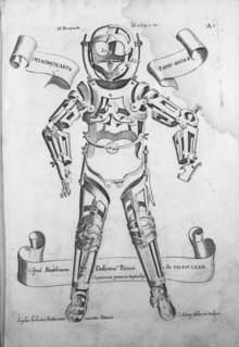
Julius Casserius (1561–1616) succeeded Fabricius as professor of anatomy at the University of Padua and published his own writings regarding technique and equipment for tracheotomy, recommending a curved silver tube with several holes in it.
Marco Aurelio Severino (1580–1656), a skillful surgeon and anatomist, performed multiple successful tracheotomies during a diphtheria epidemic in Naples in 1610, using the vertical incision technique recommended by Fabricius.
[25][26][27] Fearful of complications, most surgeons delayed the potentially life-saving tracheotomy until a patient was moribund, despite the knowledge that irreversible organ damage would have already occurred by that time.
[29] In 1871, the German surgeon Friedrich Trendelenburg (1844–1924) published a paper describing the first successful elective human tracheotomy performed to administer general anesthesia.
[30][31][32][33] After the death of German Emperor Frederick III from laryngeal cancer in 1888, Sir Morell Mackenzie (1837–1892) and the other treating physicians collectively wrote a book discussing the then-current indications for tracheotomy and when the operation is absolutely necessary.
Ironically, the newly developed inhalational anesthetic agents and techniques of general anesthesia actually seemed to increase the risks, with many patients with fatal postoperative complications.
[40] The practice of gastric endoscopy in humans was pioneered by United States Army surgeon William Beaumont (1785–1853) in 1822 with the cooperation of his patient Alexis St. Martin (1794–1880), a victim of an accidental gunshot wound to the stomach.
[41] In 1853, Antonin Jean Desormeaux (1815–1882) of Paris modified Bozzini's lichtleiter such that a mirror would reflect light from a kerosene lamp through a long metal channel.
[42][43][44][45] On 2 October 1877, Berlin urologist Maximilian Carl-Friedrich Nitze (1848–1906) and Viennese instrument maker Josef Leiter (1830–1892) introduced the first practical cystourethroscope with an electric light source.
[46] The instrument's biggest drawback was the tungsten filament incandescent light bulb (invented by Alexander Lodygin, 1847–1923), which became very hot and required a complicated water cooling system.
[57][58] In 1858, Eugène Bouchut (1818–1891), a pediatrician from Paris, developed a new technique for non-surgical orotracheal intubation to bypass laryngeal obstruction resulting from a diphtheria-related pseudomembrane.
His method involved introducing a small straight metal tube into the larynx, securing it by means of a silk thread and leaving it there for a few days until the pseudomembrane and airway obstruction had resolved sufficiently.
[60] The members of the academy rejected Bouchut's ideas, largely as a result of highly critical and negative remarks made by the influential Armand Trousseau.
[62] In November of that year, he published another study, this time on the use of orotracheal intubation to secure the airway of a patient with acute glottic edema, progressively introducing sizes 3 through 11 of "Schrotter's graduated triangular vulcanite bougies" into the larynx.
[63][64] In 1880, the Scottish surgeon William Macewen (1848–1924) reported on his use of orotracheal intubation as an alternative to tracheotomy to allow a patient with glottic edema to breathe, as well as in the setting of general anesthesia with chloroform.
[64][65][66] All previous observations of the glottis and larynx (including those of García, Hack and Macewen) had been performed under indirect vision (using mirrors) until 23 April 1895, when Alfred Kirstein (1863–1922) of Germany first described direct visualization of the vocal cords.
[72] While practicing at Bellevue Hospital in New York City, Janeway was of the opinion that direct intratracheal insufflation of volatile anesthetics would provide improved conditions for otolaryngologic surgery.
Working at the Queen's Hospital for Facial and Jaw Injuries in Sidcup with plastic surgeon Sir Harold Gillies (1882–1960) and anesthetist E. Stanley Rowbotham (1890–1979), Magill developed the technique of awake blind nasotracheal intubation.
[85] In 1949, Macintosh published a case report describing the novel use of a gum elastic urinary catheter as an endotracheal tube introducer to facilitate difficult tracheal intubation.
[86] Inspired by Macintosh's report, P. Hex Venn (who was at that time the anesthetic advisor to the British firm Eschmann Brothers & Walsh, Ltd.) set about developing an endotracheal tube introducer based on this concept.


From Angelo Mariani and Joseph Uzanne (1894). Figures contemporaines tirées de l'Album Mariani , Volume I. Paris:Ernest Flammarion.
