White blood cell
Historically, white blood cells were classified by their physical characteristics (granulocytes and agranulocytes), but this classification system is less frequently used now.
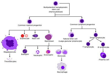
Produced in the bone marrow, white blood cells defend the body against infections and disease.
The buffy coat may sometimes be green if there are large amounts of neutrophils in the sample, due to the heme-containing enzyme myeloperoxidase that they produce.
These broadest categories can be further divided into the five main types: neutrophils, eosinophils, basophils, lymphocytes, and monocytes.
[6] A good way to remember the relative proportions of WBCs is "Never Let Monkeys Eat Bananas".
Granulocytes are distinguished from agranulocytes by their nucleus shape (lobed versus round, that is, polymorphonuclear versus mononuclear) and by their cytoplasm granules (present or absent, or more precisely, visible on light microscopy or not thus visible).
They are usually first responders to microbial infection; their activity and death in large numbers form pus.
[13] This gives the neutrophils the appearance of having multiple nuclei, hence the name polymorphonuclear leukocyte.
Neutrophils are active in phagocytosing bacteria and are present in large amount in the pus of wounds.
The average lifespan of inactivated human neutrophils in the circulation has been reported by different approaches to be between 5 and 135 hours.
They are rare in the blood, but numerous in the mucous membranes of the respiratory, digestive, and lower urinary tracts.
The most important causes of eosinophilia include allergies such as asthma, hay fever, and hives; and parasitic infections.
They secrete chemicals that destroy large parasites, such as hookworms and tapeworms, that are too big for any one white blood cell to phagocytize.
[13] The cytoplasm is full of granules that assume a characteristic pink-orange color with eosin staining.
[17] They can be recognized by several coarse, dark violet granules, giving them a blue hue.
It also makes blood vessels more permeable so neutrophils and clotting proteins can get into connective tissue more easily.
Basophils can also release chemical signals that attract eosinophils and neutrophils to an infection site.
Lymphocytes are distinguished by having a deeply staining nucleus that may be eccentric in location, and a relatively small amount of cytoplasm.
Lymphocytes include: Monocytes, the largest type of white blood cell, share the "vacuum cleaner" (phagocytosis) function of neutrophils, but are much longer lived as they have an extra role: they present pieces of pathogens to T cells so that the pathogens may be recognized again and killed.
Monocytes eventually leave the bloodstream and become tissue macrophages, which remove dead cell debris as well as attack microorganisms.
Unlike neutrophils, monocytes are able to replace their lysosomal contents and are thought to have a much longer active life.
Some leucocytes migrate into the tissues of the body to take up a permanent residence at that location rather than remaining in the blood.
Proliferative disorders of white blood cells can be classed as myeloproliferative and lymphoproliferative.
For example, the most common cause of acquired neutropenia is drug-induced, so an individual may have symptoms of medication overdose or toxicity.
Like neutropenia, symptoms and treatment of lymphocytopenia are directed at the underlying cause of the change in cell counts.
[18] Leukocytosis may affect one or more cell lines and can be neutrophilic, eosinophilic, basophilic, monocytosis, or lymphocytosis.