Mites of livestock
Other mites are parasitic, and those that infest livestock animals cause many diseases that are widespread, reduce production and profit for farmers, and are expensive to control.
Infestation by mites usually causes skin diseases known as mange, scab, demodecosis, or in general as acariasis.
The list below (Outline classification) is simplified for this veterinary article by omitting the superorders, orders, and suborders; this emphasizes the pragmatic taxonomic level of families, together with vernacular names.
However, the mites parasitic on vertebrate animals can readily be identified to at least the level of genus by nonspecialists if the clinical context of host species and site of infestation on skin or other organs is used.
Distinguishing acarines from insects (subphylum Hexapoda) is similarly important because the term 'insect' is often used in popular text and speech for various small crawling animals.
The generalized anatomy of an acarine as shown in the diagram below can be compared with that of an insect as in typical parasitic flies of domestic animals.
The early form of the female is described as pubescent (ready for mating) and may be equipped with protuberances that couple with matching sockets on the male during fertilization.
This type of lifecycle, with all active stages resembling each other in structure and feeding mechanism, is called incomplete metamorphosis (or hemimetabolism).
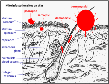
[9] The position of these mites in relation to the skin or other organs is crucial for their feeding and also for their veterinary diagnosis and options for treatment.
[12] Demodectic mites have mouthparts adapted to penetrate and suck out the contents of individual cells of hair follicles.
The tube through which food is ingested and saliva excreted during feeding is formed in most mites by apposing the sheath that contains the chelicerae against the hypostome.
[20][21] The lifecycle starts with eggs laid in the environment of the normal hosts of the larvae, typically rodents and other small mammals.
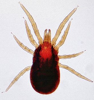
They actively hunt amongst the ground-level debris layer of vegetation and are conspicuous by their red color and dense outer covering of fine setae, appearing like fur.
[22] These mites remain external to the true outer layer of the skin (the epidermis) which also lines the tube of the hair follicle.
Demodex mites are morphologically adapted to this constricted habitat: microscopic, worm shaped, and with very short legs.
[9] They have long, powerful legs that they use to seek their hosts and their size and active crawling cause them to resemble larval ticks.
Irritation of the outer skin by the mite's mouthparts and saliva results in a complex form of cutaneous hypersensitivity and inflammatory exudation of serum and fresh cells.
[3] In contrast, Psorergates ovis feeding on sheep induces inflammatory and hypersensitive reactions in the epidermis, resulting in intense pruritus and formation of scabs.
[30] The females, after mating with males on the surface of their host's skin, burrow into the living layers of the epidermis (mainly the stratum spinosum).
Chronic infestations lead to thickening of the skin by overproduction of epidermal cells (acanthosis), resulting in a characteristic depilated and scaly appearance.
[3] Demodecosis in cattle caused by Demodex bovis can occur as dense, localized infestations with inflammation of the epidermis in hair follicles.
Eutrombicula alfreddugesi and E. splendens are common in the Americas where they infest the skin of the face, muzzle, legs, and belly of cattle and horses.
(Trombiculid mites also leave intensely pruritic spots on dogs and humans after feeding and such infestation is closely associated with the grassland and scrub habitats of the nonparasitic stages of the lifecycle.)
Originally a parasite centered on the nests of its wild bird hosts, this species has become a major pest in commercial poultry houses.
Dermanyssid mites are also implicated in the transmission to poultry birds of the bacterial agent of avian spirochaetosis, Borrelia anserina, but the main transmitters of this bacterium are argasid ticks.
(Trombiculid mites are notorious as transmitters of Orientia tsutsugamushi, the bacterium that causes scrub typhus in humans in Southeast Asia.
However, alternatives such as hand-held sprayers and pour-on applicators are now often used to reduce costs and risk of contamination associated with running a dip tank.
[44] A more direct way of treating some types of mite are antiparasitic drugs that act systemically through the internal circulation of the animal rather than topically on the skin.
[47] Fungi such as Metarhizium anisopliae and Beauveria bassiana that are naturally pathogenic to mites are subjects of research as an alternative to chemical pesticides.
Self-dusting by birds is a natural defense against mites (and lice) and can be aided by poultry farmers providing diatomaceous earth (also known as kaolin).