Monckeberg's arteriosclerosis
Calcium deposits are found in the muscular middle layer of the walls of arteries (the tunica media)[1] with no obstruction of the lumen.
Stage 1 involves the formation of calcium deposits both inside and outside the vascular smooth muscle cells which compose the tunica media.
Calcification outside of the vascular smooth muscle cells are commonly associated with damage to elastic fibers in the extra-cellular matrix.
[8] Monckeberg's calcification typically occurs near the internal elastic lamina or, less frequently, in the media of muscular arteries without alterations in calcium metabolism.
However, another study, which examined 143 histologically normal femoral arteries from young, healthy multi-organ donors, suggests otherwise.
This indicates that Monckeberg's calcification may develop early in life, potentially due to abnormal osteogenic differentiation of vascular progenitor cells.
Intercranial Mönckeberg's arteriosclerosis has been commonly found in people with malignant tumors who died of cerebral infarctions.
[16] Mönckeberg's arteriosclerosis is typically an incidental finding, detected through clinical examination or plain radiography, and may be associated with diabetes mellitus or chronic kidney disease.
[19][20] In addition, elevated inorganic phosphate may have direct signaling effects which can induce the progression of Mönckeberg's arteriosclerosis.
[3] However, studies in animals suggest that there is mainly a medial pattern of vascular calcification reflects different underlying mechanisms of disease,[17] and despite involvement of the internal elastic lamina, evidence of inflammation is rare in Mönckeberg's arteriosclerosis.
[28][29] However this type of non-invasive diagnostic tool could lead to falsely elevated values, especially individuals with diabetes that have lower limb ischemia.
In an observational study, 11% of patients that met the criteria of diabetes and critical ischemia had exhibited false ABI levels.
Lowering calcium and phosphate levels in people with calciphylaxis, along with increasing hemodialysis and treating potential ischemic necrosis is also recommended.
Some promising studies have been exploring this concept and have created 3D printed tubular structures similar to the human body's own vasculature to use as a model for testing.
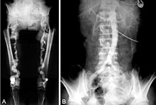
[33] A 28-year-old male in Saudi Arabia presented with swelling in both of his thighs, despite no past medical history significant of chronic illness, trauma, or surgeries.
Extensive analysis of the individual's condition lead medical professionals to the conclusion that Mönckeberg's arteriosclerosis was suspected as the cause for their symptoms.
[26] A 62-year-old diabetic male presented with angina that had lasted for six days until coming in for a coronary angiography when it was noticed they had signs of Mönckeberg's arteriosclerosis.
All routine labs of the individual were at normal levels but it was noticed that the patient may have recently experienced a myocardial infarction.
The individual had their pain, which was produced by Mönckeberg medial arteriosclerosis, controlled by using high dosed corticosteroids combined with the usage of warm compresses and transcutaneous electrical nerve stimulation (TENS).
[35] A case report describes a 69-year-old female with Monckeberg’s arteriosclerosis affecting her uterine vessels, following long-term endometritis and experiencing premature menopause.
[3] The prevalence of Mönckeberg's arteriosclerosis in the general population has been estimated as <1% on the basis of an ankle brachial pressure index >1.5;[29][38][39] however the validity of this criterion is questionable.