Myogenesis
Studies have shown that even rat and chick myoblasts can recognise and align with one another, suggesting evolutionary conservation of the mechanisms involved.
Fusion involves recruitment of actin to the plasma membrane, followed by close apposition and creation of a pore that subsequently rapidly widens.
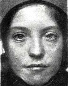
[3] Waardenburg syndrome is most often associated with congenital disorders involving the intestinal tract and spine, an elevation of the scapula, among other symptoms.
[3] Associated Genetic Factors: PAX3, c-Met, Mox2, MSX1, Six, Myf5, and MyoD Mox2 (also referred to as MEOX-2) plays an important role in the induction of mesoderm and regional specification.
[3] Impairing the function of Mox2 will prevent the proliferation of myogenic precursors and will cause abnormal patterning of limb muscles.
[3] Studies have shown that mice muscle development in the intercostal and paraspinal regions can be delayed by inactivating Myf-5.
[3] These consequences further reveal the complexity of myogenesis and the importance of each genetic factor in proper muscle development.
Both MyoD and Myf5 are members of the myogenic bHLH (basic helix-loop-helix) proteins transcription factor family.
[6] Consequently, the simultaneous deletion of Myf5 and MyoD also results in a complete lack of skeletal muscle formation.
The consequences of this abnormal patterning include severe reduction in size of hindlimbs and complete absence of forelimb muscles.
Activated by stimuli such as injury or high mechanical load, satellite cells are required for muscle regeneration in adult organisms.
[3] During embryogenesis, the dermomyotome and/or myotome in the somites contain the myogenic progenitor cells that will evolve into the prospective skeletal muscle.
[9] The determination of dermomyotome and myotome is regulated by a gene regulatory network that includes a member of the T-box family, tbx6, ripply1, and mesp-ba.
[10] Skeletal myogenesis depends on the strict regulation of various gene subsets in order to differentiate the myogenic progenitors into myofibers.
[9] MRF4 is important for blocking the transcription of muscle-specific promoters, enabling skeletal muscle progenitors to grow and proliferate before differentiating.
[5] The O subfamily of the forkhead proteins (FOXO) play a critical role in regulation of myogenic differentiation as they stabilize Notch/Hes binding.
Research has shown that knockout of FOXO1 in mice increases MyoD expression, altering the distribution of fast-twitch and slow-twitch fibers.
[14] In myoblasts, PtdIns5P, produced by the lipid phosphatase MTM1, is rapidly metabolized by PI5P 4-kinase α into PI(4,5)P2, which accumulates at the plasma membrane.
This accumulation facilitates the formation of podosome-like protrusions, where the fusogen Myomaker is localized, playing a crucial role in the spatiotemporal regulation of myoblast fusion.
It accomplishes this by repressing MyoD transcription by binding to the enhancer region, and prevents premature myogenesis.
Gain and loss of this ligand in neural crest cells results in delayed or premature myogenesis.
[9] This approach, using cell based high-throughput transfection assay and whole-mount in situ hybridization, was used in identifying the myogenetic regulator RP58, and the tendon differentiation gene, Mohawk homeobox.