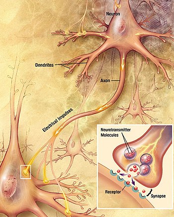
Neuron
Molecular evidence suggests that the ability to generate electric signals first appeared in evolution some 700 to 800 million years ago, during the Tonian period.
They eventually gained new gene modules which enabled cells to create post-synaptic scaffolds and ion channels that generate fast electrical signals.
Motor neurons receive signals from the brain and spinal cord to control everything from muscle contractions[3] to glandular output.
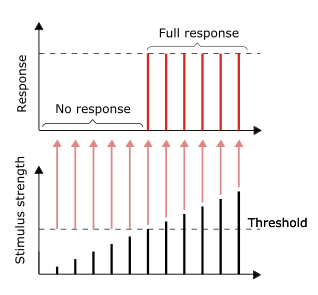
If the voltage changes by a large enough amount over a short interval, the neuron generates an all-or-nothing electrochemical pulse called an action potential.
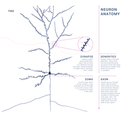
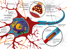
Neurons are the primary components of the nervous system, along with the glial cells that give them structural and metabolic support.
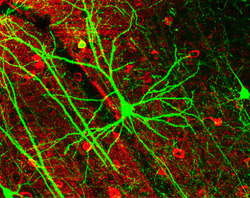
Given the diversity of functions performed in different parts of the nervous system, there is a wide variety in their shape, size, and electrochemical properties.
Sensory neurons can have axons that run from the toes to the posterior column of the spinal cord, over 1.5 meters in adults.
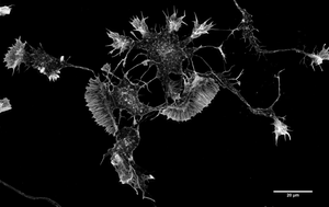
Astrocytes are star-shaped glial cells that have been observed to turn into neurons by virtue of their stem cell-like characteristic of pluripotency.
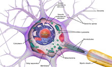
Named after German psychiatrist and neuropathologist Franz Nissl (1860–1919), they are involved in protein synthesis and their prominence can be explained by the fact that nerve cells are very metabolically active.
Basophilic dyes such as aniline or (weakly) hematoxylin[13] highlight negatively charged components, and so bind to the phosphate backbone of the ribosomal RNA.
[27] In 2023, a comprehensive cell atlas of the adult, and developing human brain at the transcriptional, epigenetic, and functional levels was created through an international collaboration of researchers using the most cutting-edge molecular biology approaches.
[38] Neurons also communicate with microglia, the brain's main immune cells via specialized contact sites, called "somatic junctions".
[39] In 1937 John Zachary Young suggested that the squid giant axon could be used to study neuronal electrical properties.
The cell membrane of the axon and soma contain voltage-gated ion channels that allow the neuron to generate and propagate an electrical signal (an action potential).
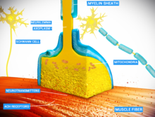
The sheath enables action potentials to travel faster than in unmyelinated axons of the same diameter, whilst using less energy.
The myelin sheath in peripheral nerves normally runs along the axon in sections about 1 mm long, punctuated by unsheathed nodes of Ranvier, which contain a high density of voltage-gated ion channels.
Greater intensity of stimulation, like brighter image/louder sound, does not produce a stronger signal but can increase firing frequency.
The neurons of the skin and muscles that are responsive to pressure and vibration have filtering accessory structures that aid their function.
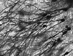
[50][48] Some previous works used nerve cell (cellule nervose), as adopted in Camillo Golgi's 1873 paper on the discovery of the silver staining technique used to visualize nervous tissue under light microscopy.
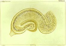
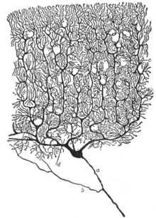
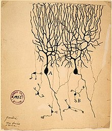
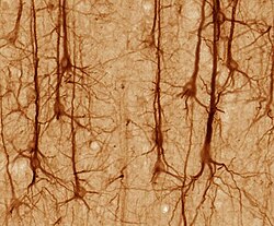
[51] The neuron's place as the primary functional unit of the nervous system was first recognized in the late 19th century through the work of the Spanish anatomist Santiago Ramón y Cajal.
[52] To make the structure of individual neurons visible, Ramón y Cajal improved a silver staining process that had been developed by Camillo Golgi.
In this paper, he stated that he could not find evidence for anastomosis between axons and dendrites and called each nervous element "an autonomous canton.
Dendritic branches can be modeled as spatial compartments, whose activity is related to passive membrane properties, but may also be different depending on input from synapses.
Compartmental modelling of dendrites is especially helpful for understanding the behavior of neurons that are too small to record with electrodes, as is the case for Drosophila melanogaster.
The fruit fly Drosophila melanogaster, a common subject in biological experiments, has around 100,000 neurons and exhibits many complex behaviors.
Many properties of neurons, from the type of neurotransmitters used to ion channel composition, are maintained across species, allowing scientists to study processes occurring in more complex organisms in much simpler experimental systems.
Charcot–Marie–Tooth disease (CMT) is a heterogeneous inherited disorder of nerves (neuropathy) that is characterized by loss of muscle tissue and touch sensation, predominantly in the feet and legs extending to the hands and arms in advanced stages.
The primary symptoms are the results of decreased stimulation of the motor cortex by the basal ganglia, normally caused by the insufficient formation and action of dopamine, which is produced in the dopaminergic neurons of the brain.
Myasthenia gravis is a neuromuscular disease leading to fluctuating muscle weakness and fatigability during simple activities.
The disintegration is dependent on ubiquitin and calpain proteases (caused by the influx of calcium ions), suggesting that axonal degeneration is an active process that produces complete fragmentation.

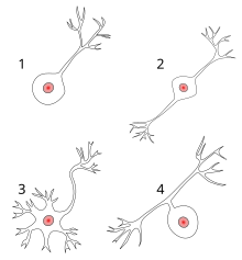
1 Unipolar neuron
2 Bipolar neuron
3 Multipolar neuron
4 Pseudounipolar neuron