Niacin test
[3] The niacin test is typically only conducted on slow-growing, granular, tan colored colonies, as these are the morphology characteristics of M. tuberculosis on an agar plate.
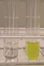
This chemical will break apart the pyridine ring of niacin to produce y-carboxy glutaconic aldehyde and joins an aromatic amine to form a yellow color.
Water is placed onto the culture plate and touched with a test strip for 15–20 minutes inside a small, sterile tube.
If excess amounts of niacin are detected, the liquid inside the tube will turn yellow, a positive test.
[5] Because lab samples that are determined to be acid-fast bacilli are possibly M. tuberculosis, a biosafety level 3 organism, all niacin tests must be conducted in a biosafety cabinet with a full gown, respirator, gloves, and sealed laboratory to ensure the safety of the laboratory technician performing the test.