Phosphoinositide phospholipase C
At present, the family consists of six sub-families comprising a total of 13 separate isoforms that differ in their mode of activation, expression levels, catalytic regulation, cellular localization, membrane binding avidity and tissue distribution.
All are capable of catalyzing the hydrolysis of PIP2 into two important second messenger molecules, which go on to alter cell responses such as proliferation, differentiation, apoptosis, cytoskeleton remodeling, vesicular trafficking, ion channel conductance, endocrine function and neurotransmission.
All family members are capable of catalyzing the hydrolysis of PIP2, a phosphatidylinositol at the inner leaflet of the plasma membrane into the two second messengers, inositol trisphosphate (IP3) and diacylglycerol (DAG).
However, in the second phosphodiesterase step, the cyclic intermediate is held within the active site long enough to be attacked by a molecule of water, resulting in a final acyclic IP3 product.
It should be mentioned that bacterial forms of the enzyme, which contain only the catalytic lipase domain, produce cyclic intermediates exclusively, whereas the mammalian isoforms generate predominantly the acyclic product.
It is important to note that research has also discovered that, in addition to the plasma membrane, phosphoinositide phospholipase C also exists within other sub-cellular regions such as the cytoplasm and nucleus of the cell.
The β subfamily is distinguished from the others by the presence of a long C-terminal extension immediately downstream of the C2 domain, which is required for activation by Gαq subunits, and which plays a role in plasma membrane binding and nuclear localization.
Members of the Rho GTPase family (e.g., Rac1, Rac2, Rac3, and cdc42) have been implicated in their activation by binding to an alternate site on the N-terminal PH domain followed by subsequent recruitment to the plasma membrane.
A basic amino acid region within the enzyme's long C-terminal tail appears to function as a Nuclear Localization Signal for import into the nucleus.
PLC-γ2 then competes with PI-3K for PIP2 which it hydrolyzes into IP3 (inositol 1,4,5-trisphosphate), which ultimately raises intercellular calcium, and diacylglycerol (DAG), which activates portions of the PKC family.
In addition, this specificity helps tether the enzyme tightly to the plasma membrane in order to access substrate through ionic interactions between the phosphate groups of PIP2 and charged residues in the PH domain.
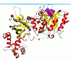
The widely expressed PLC-δ1 isoform is the best-characterized phospholipase family member, as it was the first to have high-resolution X-ray crystal structures available for analysis.
In terms of domain architecture, all of the enzymes are built upon a common PLC-δ backbone, wherein each family displays similarities, as well as obvious distinctions, that contribute to unique regulatory properties within the cell.
Because it is the only family found expressed in lower eukaryotic organisms such as yeast and slime molds, it is considered the prototypical PLC isoform.