Sphingomyelin
It usually consists of phosphocholine and ceramide, or a phosphoethanolamine head group; therefore, sphingomyelins can also be classified as sphingophospholipids.
[1][2] In humans, SPH represents ~85% of all sphingolipids, and typically makes up 10–20 mol % of plasma membrane lipids.
Sphingomyelin obtained from natural sources, such as eggs or bovine brain, contains fatty acids of various chain length.
[6] Sphingomyelin is synthesized at the endoplasmic reticulum (ER), where it can be found in low amounts, and at the trans Golgi.
[7] The Golgi complex represents an intermediate between the ER and plasma membrane, with slightly higher concentrations towards the trans side.
[7] The phosphocholine head group is released into the aqueous environment while the ceramide diffuses through the membrane.
In addition, the degradation of sphingomyelin can produce ceramide which is involved in the apoptotic signaling pathway.
Studies in the late 1990s had found that ceramide was produced in a variety of conditions leading to apoptosis.
[14] It was then hypothesized that sphingomyelin hydrolysis and ceramide signaling were essential in the decision of whether a cell dies.
In the early 2000s new studies emerged that defined a new role for sphingomyelin hydrolysis in apoptosis, determining not only when a cell dies but how.
[14] After more experimentation it has been shown that if sphingomyelin hydrolysis happens at a sufficiently early point in the pathway the production of ceramide may influence either the rate and form of cell death or work to release blocks on downstream events.
This order is in part due to the higher transition temperature of sphingolipids as well as the interactions of these lipids with cholesterol.
Cholesterol is a relatively small, nonpolar molecule that can fill the space between the sphingolipids that is a result of the large acyl chains.
A hemolytic protein, lysenin, may be a valuable probe for sphingomyelin detection in cells of Niemann-Pick A patients.
MS patients exhibit upregulation of certain cytokines in the cerebrospinal fluid, particularly tumor necrosis factor alpha.

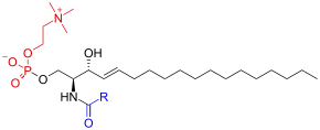
Black: Sphingosine
Red: Phosphocholine
Blue: Fatty acid

