Thymoma
These problems may take the form of superior vena cava syndrome, dysphagia (difficulty swallowing), cough, or chest pain.
Other reported disease associations are with acute pericarditis, agranulocytosis, alopecia areata, ulcerative colitis, Cushing's disease, hemolytic anemia, limbic encephalopathy, myocarditis, nephrotic syndrome, panhypopituitarism, pernicious anemia, polymyositis, rheumatoid arthritis, sarcoidosis, scleroderma, sensorimotor radiculopathy, stiff person syndrome, systemic lupus erythematosus and thyroiditis.
[2][4] One-third to one-half of all persons with thymoma have no symptoms at all, and the mass is identified on a chest X-ray or CT/CAT scan performed for an unrelated problem.
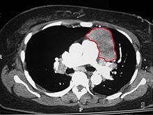
When a thymoma is suspected, a CT/CAT scan is generally performed to estimate the size and extent of the tumor, and the lesion is sampled with a CT-guided needle biopsy.
These include: full blood count, protein electrophoresis, antibodies to the acetylcholine receptor (indicative of myasthenia), electrolytes, liver enzymes and renal function.
If the tumor is apparently invasive and large, preoperative (neoadjuvant) chemotherapy and/or radiotherapy may be used to decrease the size and improve resectability, before surgery is attempted.
Invasive thymomas uncommonly can also metastasize, generally to pleura, bones, liver or brain in approximately 7% of cases.
[2] A study found that slightly over 40% of observed patients with stage III and IV tumors survived for at least 10 years after diagnosis.