[5] Tyrosinase is a copper-containing enzyme present in plant and animal tissues that catalyzes the production of melanin and other pigments from tyrosine by oxidation.
The two copper atoms within the active site of tyrosinase enzymes interact with dioxygen to form a highly reactive chemical intermediate that then oxidizes the substrate.
Tyrosinases and catechol oxidases are collectively termed polyphenol oxidases.Tyrosinases have been isolated and studied from a wide variety of plant, animal, and fungal species.
Tyrosinases from different species are diverse in terms of their structural properties, tissue distribution, and cellular location.
[10] The enzymes found in plant, animal, and fungal tissue frequently differ with respect to their primary structure, size, glycosylation pattern, and activation characteristics.
It is most common in Europe, but is also found at lower, moderate frequencies in Central Asia, the Middle East, North Africa, and among the San and Mbuti Pygmies.
Decreasing tyrosinase activity has been targeted for the improvement or prevention of conditions related to the hyperpigmentation of the skin, such as melasma and age spots.
[22] Several polyphenols, including flavonoids or stilbenoid, substrate analogues, free radical scavengers, and copper chelators, have been known to inhibit tyrosinase.
[28] Tyrosinase family related genes plays an important role in the evolution, genetics, and developmental biology of pigment cells, as well as to approach human disorders associated with defects in their synthesis, regulation or function in vertebrates three types of melanin producing pigment cells are well known since embryonic origin i.e., from the neural crest, neural tube and pineal body.
Their biosynthesis is governed by evolutionary conserved enzymes of the tyrosinase family( tyr, tyr1 and tyr2) also called DOPAchrome tautomerase (dct).
[29] Similarly, the type-3 copper protein family perform various biological function including pigment formation, innate immunity and oxygen transport.
The combine genetic phylogenetic and structural analysis concluded that the original type-3 copper protein possessed a single peptide and grouped into α subclass.
The ancestral protein gene underwent to two duplication i.e., first one prior to divergence of unknown eukaryotic lineage and second one before diversification.
Recent research by cosmetic companies has been focused on the development of novel whitening agents that selectively suppress tyrosinase activity to reduce hyperpigmentation while avoiding cytotoxicity of healthy melanocytes.
[36] Traditional pharmacological agents such as corticosteroids, hydroquinone, and amino numeric chloride lighten skin through the inhibition of melanocyte maturation.
Tyrosinase has a wide range of functions in insects, including wound healing, sclerotization, melanin synthesis and parasite encapsulation.
Crystallographic structure
of a
Streptomyces
-derived tyrosinase in complex with a so-called "caddie protein".
[
8
]
In all models, only the tyrosinase molecule is shown, copper atoms are shown in green and the molecular surface is shown in red. In models D and E, histidine amino acids are shown as a blue line representation. From model E, each copper atom within the active site is indeed complexed with three
histidine
residues, forming a
type 3 copper center
. From models C and D, the active site for this protein can be seen to sit within a pillus formed on the molecular surface of the molecule.
This is an alignment structure showing only the conserved region of protein nucleotide sequences of Frogs (Their genebank accession number
CAR95491
,
CAJ82935
,
BAA02077
,
BAV78831
and
AAC17168
), Snakes (Their genebank accession numbers
BBC55580
,
XP032076040
and
BBC55647
) and Human (Genebank accession number
AAA61242
) using Clustal Omega. (Note: (*) shows a conserved region,(.) shows more conserved and (:) shows less conserved.)
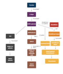
The Raper–Mason (melanogenesis) pathway, depicting the steps in melanin synthesis
[
20
]
.
DHI- 5,6 dihydroxyindole,
DHICA
- 5,6- dihydroxyphenylalanine,
GGT
- Gamma-glutamyl transpeptidase,
GST
- Glutathione-S-transferase;
L-Dopa
- Levo- Dopa,
TRP-2
- Tyrosinase- related protein 2
A representative
phylogenetic tree
cladogram
of tyrosinase proteins. Tyrosinase sequences from ten vertebrates species( Genus:
Ambystoma, Xenopus, Homo, Elaphe, Thamnophis, Bufo, Rugosa, and Rana)
were analyzed. The multiple alignments are generated by the
CLUSTAL W
program( version 1.7)and the phylogenetic trees were constructed by the Neighbour-joining method without distance correction. So
Ambystoma and Xenopus
do not cluster with other amphibians. Branches and nodes are drawn according to identical patterns.
Mutations in the tyrosinase gene that have been shown to cause albinism in animals. Colored boxes indicate regions in the protein encoded by one of five exons (see figure of gene structure). Positions refer to amino acid positions in protein of each species. Modified after Miura et al.
Substituted amino acids of tyrosinase in albino frogs and corresponding amino acids in other vertebrate species (Miura et al., 2018). kW refers to the rice frog
kawamurai
Wild type, kA: kawamurai Albino type, rW: rugosa Wild, rA:
rugosa
Albino, nW
nigromaculatus
Wild, nA
H
: nigromaculatus Albino collected from Hiroshima. Numbers outside of the parenthesis refer to the amino acid position of the mutated species, and the number in the parenthesis refers to the associated amino acid position in the human sequence. (Miura et al. 2018)
[
26
]
This is a schematic representation of the intron-exon organization of tyrosinase (TYP) gene in humans (ClinVar:
NM_ 000372
).
[
27
]
Open and closed boxes represent protein-coding and untranslated regions of exons, respectively, with exons labeled by numbers. Intron sizes are indicated by small numbers (in bp).
ConSurf
uses a series of nine colors from turquoise through white through
burgundy
to represent conservation grades from variable through conserved, respectively. At right is an alternative color scheme inspired by the earlier (now obsolete) ProteinExplorer's MSA3D (in which grades 4, 5, and 6 use the same color).