Visual system
The system detects, transduces and interprets information concerning light within the visible range to construct an image and build a mental model of the surrounding environment.
The visual system performs a number of complex tasks based on the image forming functionality of the eye, including the formation of monocular images, the neural mechanisms underlying stereopsis and assessment of distances to (depth perception) and between objects, motion perception, pattern recognition, accurate motor coordination under visual guidance, and colour vision.
V2 serves much the same function as V1, however, it also handles illusory contours, determining depth by comparing left and right pulses (2D images), and foreground distinguishment.
[17][18] The inferior temporal gyrus recognizes complex shapes, objects, and faces or, in conjunction with the hippocampus, creates new memories.
[20] The Edinger-Westphal nucleus moderates pupil dilation and aids (since it provides parasympathetic fibers) in convergence of the eyes and lens adjustment.
[25] There are three types of cones that differ in the wavelengths of light they absorb; they are usually called short or blue, middle or green, and long or red.
Cones mediate day vision and can distinguish color and other features of the visual world at medium and high light levels.
In the dark, the chromophore retinal has a bent shape called cis-retinal (referring to a cis conformation in one of the double bonds).
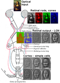
[27][28] The final result of all this processing is five different populations of ganglion cells that send visual (image-forming and non-image-forming) information to the brain:[26] A 2006 University of Pennsylvania study calculated the approximate bandwidth of human retinas to be about 8,960 kilobits per second, whereas guinea pig retinas transfer at about 875 kilobits.
[29] In 2007 Zaidi and co-researchers on both sides of the Atlantic studying patients without rods and cones, discovered that the novel photoreceptive ganglion cell in humans also has a role in conscious and unconscious visual perception.
The functioning of a camera is often compared with the workings of the eye, mostly since both focus light from external objects in the field of view onto a light-sensitive medium.
With this simple geometrical similarity, based on the laws of optics, the eye functions as a transducer, as does a CCD camera.
In the visual system, retinal, technically called retinene1 or "retinaldehyde", is a light-sensitive molecule found in the rods and cones of the retina.
This parallel processing is important for reconstructing the visual world; each type of information will go through a different route to perception.
Another population sends information to the superior colliculus in the midbrain, which assists in controlling eye movements (saccades)[31] as well as other motor responses.
A final population of photosensitive ganglion cells, containing melanopsin for photosensitivity, sends information via the retinohypothalamic tract to the pretectum (pupillary reflex), to several structures involved in the control of circadian rhythms and sleep such as the suprachiasmatic nucleus (the biological clock), and to the ventrolateral preoptic nucleus (a region involved in sleep regulation).
[32] A recently discovered role for photoreceptive ganglion cells is that they mediate conscious and unconscious vision – acting as rudimentary visual brightness detectors as shown in rodless coneless eyes.
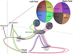
[26] The optic radiations, one on each side of the brain, carry information from the thalamic lateral geniculate nucleus to layer 4 of the visual cortex.
In the parietal lobe, the lateral and ventral intraparietal cortex are involved in visual attention and saccadic eye movements.
Pediatricians are able to perform non-verbal testing to assess visual acuity of a newborn, detect nearsightedness and astigmatism, and evaluate the eye teaming and alignment.
From recent studies in the United States and Australia there is some evidence that the amount of time school aged children spend outdoors, in natural light, may have some impact on whether they develop myopia.
A number of changes occur with aging: Along with proprioception and vestibular function, the visual system plays an important role in the ability of an individual to control balance and maintain an upright posture.
Difficulty in sensing, processing and understanding light input has the potential to adversely impact an individual's ability to communicate, learn and effectively complete routine tasks on a daily basis.
In children, early diagnosis and treatment of impaired visual system function is an important factor in ensuring that key social, academic and speech/language developmental milestones are met.
This typically involves the outer layers of the optic nerve, sometimes as a result of buildup of fluid and excessive pressure in the eye.
The variety of cones enables them to perceive an enhanced array of colors as a mechanism for mate selection, avoidance of predators, and detection of prey.
[58] Certain one-celled microorganisms, the warnowiid dinoflagellates have eye-like ocelloids, with analogous structures for the lens and retina of the multi-cellular eye.
[59] The armored shell of the chiton Acanthopleura granulata is also covered with hundreds of aragonite crystalline eyes, named ocelli, which can form images.
[60] Many fan worms, such as Acromegalomma interruptum which live in tubes on the sea floor of the Great Barrier Reef, have evolved compound eyes on their tentacles, which they use to detect encroaching movement.
[62] Biologists have determined that humans have extremely good vision compared to the overwhelming majority of animals, particularly in daylight, surpassed only by a few large species of predatory birds.


The image projected onto the retina is inverted due to the optics of the eye.

V1; V2; V3; V4; V5 (also called MT)

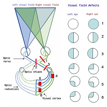
From top to bottom:
1. Complete loss of vision, right eye
2. Bitemporal hemianopia
3. Homonymous hemianopsia
4. Quadrantanopia
5&6. Quadrantanopia with macular sparing
