Visual cycle
Retinal is the chromophore of most visual opsins, meaning it captures the photons to begin the phototransduction cascade.
Each molecule of retinal must travel from the photoreceptor cell to the RPE and back in order to be refreshed and combined with another opsin.
Retinal can be photoisomerized by itself, but requires to be bound to an opsin protein to both trigger the phototransduction cascade and tune the spectral sensitivity to longer wavelengths, which enable color vision.
To reach the retina, it is bound to Retinol Binding Protein (RBP) and Transthyretin, which prevents its filtration in the glomeruli.
It then proceeds to the cell membrane of the rod, where it is chaperoned to the Retinal Pigment Epithelium (RPE) by Interphotoreceptor retinoid-binding protein (IRBP).
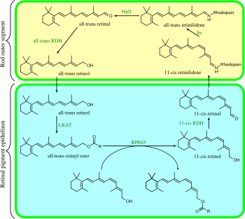
When inside the RPE cell, bound to CRBP, the all-trans-retinol is esterified by Lecithin Retinol Acyltransferase (LRAT) to form a retinyl ester.
When further chromophore is required, the retinyl esters are acted on by isomerohydrolase to produce 11-cis-retinol, which is transferred to the Cellular retinaldehyde binding protein (CRALBP).
It is believed that an alternative visual cycle exists, which uses Müller glial cells instead of Retinal Pigment Epithelium.
However, unlike the rod and cone pigments, melanopsin has the ability to act as both the excitable photopigment and as a photoisomerase.
As a result, the rods continually secrete glutamate, a neurotransmitter, at a rate the Muller cells are unable to absorb.