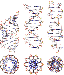
A-DNA
However, as shown in the comparison table below, there is a slight increase in the number of base pairs (bp) per turn.
[2] These intermediates can form in aqueous solutions when the cytosine bases are methylated or brominated, altering the conformation.
Alternatively, guanine and cytosine rich fragments have been shown to be easily converted from B to A-form in aqueous solutions.
[5][1] Protein binding can also strip solvent off of DNA and convert it to the A form, as revealed by the structure of several hyperthermophilic archaeal viruses.
These viruses include rod-shaped rudiviruses SIRV2 [6] and SSRV1,[7] enveloped filamentous lipothrixviruses AFV1,[8] SFV1 [9] and SIFV,[7] tristromavirus PFV2 [10] as well as icosahedral portoglobovirus SPV1.