Angiography
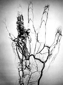
Angiography or arteriography is a medical imaging technique used to visualize the inside, or lumen, of blood vessels and organs of the body, with particular interest in the arteries, veins, and the heart chambers.
Modern angiography is performed by injecting a radio-opaque contrast agent into the blood vessel and imaging using X-ray based techniques such as fluoroscopy.
The technique was first developed in 1927 by the Portuguese physician and neurologist Egas Moniz at the University of Lisbon to provide contrasted X-ray cerebral angiography in order to diagnose several kinds of nervous diseases, such as tumors, artery disease and arteriovenous malformations.
For example, in 1932, Lopo de Carvalho performed the first pulmonary angiogram via venous puncture of the superior member.
With the introduction of the Seldinger technique in 1953, the procedure became markedly safer as no sharp introductory devices needed to remain inside the vascular lumen.
Both these techniques enable the interventional radiologist or cardiologist to see stenosis (blockages or narrowings) inside the vessel which may be inhibiting the flow of blood and causing pain.
After the procedure has been completed, if the femoral technique is applied, the site of arterial entry is either manually compressed, stapled shut, or sutured in order to prevent access-site complications.
A long, thin, flexible tube called a catheter is used to administer the X-ray contrast agent at the desired area to be visualized.
[6] Cerebral angiography provides images of blood vessels in and around the brain to detect abnormalities, including arteriovenous malformations and aneurysms.
[11] Angiography is also commonly performed to identify vessels narrowing in patients with leg claudication or cramps, caused by reduced blood flow down the legs and to the feet; in patients with renal stenosis (which commonly causes high blood pressure) and can be used in the head to find and repair stroke.
[14] Optical coherence tomography (OCT) is a technology using near-infrared light to image the eye, in particular penetrate the retina to view the micro-structure behind the retinal surface.
Post mortem CT angiography for medicolegal cases is a method initially developed by a virtopsy group.
The risk of complications from angiography can be reduced with a prior CT scan by providing clinicians with more information about number and positioning of the clots in advance.
[19][20] Major complications in cerebral angiography such as in digital subtraction angiography or contrast MRI are also rare but include stroke, an allergic reaction to the anaesthetic other medication or the contrast medium, blockage or damage to one of the access veins in the leg, pseudoaneurysm at the puncture site; or thrombosis and embolism formation.
[21] The contrast medium that is used usually produces a sensation of warmth lasting only a few seconds, but may be felt in a greater degree in the area of injection.
If digital subtraction angiography is used instead, the risks are considerably reduced because the catheter does not need to be passed as far into the blood vessels; thus lessening the chances of damage or blockage.