Atherosclerosis
[24] Marked narrowing in the coronary arteries, which are responsible for bringing oxygenated blood to the heart, can produce symptoms such as chest pain of angina and shortness of breath, sweating, nausea, dizziness or lightheadedness, breathlessness or palpitations.
Stroke is caused by marked narrowing or closure of arteries going to the brain; lack of adequate blood supply leads to the death of the cells of the affected tissue.
[26] Peripheral arteries, which supply blood to the legs, arms, and pelvis, also experience marked narrowing due to plaque rupture and clots.
[55] Rats fed DHA-containing oils experienced marked disruptions to their antioxidant systems, and accumulated significant amounts of phospholipid hydroperoxide in their blood, livers and kidneys.
[57] In another study, rabbits fed heated soybean oil "grossly induced atherosclerosis and marked liver damage were histologically and clinically demonstrated.
One recent hypothesis suggests that, for unknown reasons, leukocytes, such as monocytes or basophils, begin to attack the endothelium of the artery lumen in cardiac muscle.
Chronic inflammation within the arterial wall, driven by immune cells like macrophages, accelerates atherosclerotic plaque instability by promoting collagen breakdown and thinning the fibrous cap, which increases the likelihood of rupture and thrombosis.
[65] Autopsy studies have shown that the prevalence of coronary artery atherosclerosis in males from the United States, with an average age of 22.1 years old, who died in war, ranges from 45% to 77.3%.
[9][67] The primary documented driver of this process is oxidized lipoprotein particles within the wall, beneath the endothelial cells, though upper normal or elevated concentrations of blood glucose also plays a major role and not all factors are fully understood.
[citation needed] Low-density lipoprotein (LDL) particles in blood plasma invade the endothelium and become oxidized, creating risk of cardiovascular disease.
[citation needed] In addition to these cellular activities, there is also smooth muscle proliferation and migration from the tunica media into the intima in response to cytokines secreted by damaged endothelial cells.
A similar form of intramural calcification, presenting the picture of an early phase of arteriosclerosis, appears to be induced by many drugs that have an antiproliferative mechanism of action (Rainer Liedtke 2008).
To attract and stimulate macrophages, the cholesterol must be released from the LDL particles and oxidized, a key step in the ongoing inflammatory process.
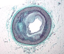
As long as the artery enlarges sufficiently to compensate for the extra thickness of the atheroma, then no narrowing ("stenosis") of the opening ("lumen") occurs.
In addition, the calcification deposits between the outer portion of the atheroma and the muscular wall, as they progress, lead to a loss of elasticity and stiffening of the artery as a whole.
Stenoses can be slowly progressive, whereas plaque ulceration is a sudden event that occurs specifically in atheromas with thinner/weaker fibrous caps that have become "unstable".
[citation needed] If the fibrous cap separating a soft atheroma from the bloodstream within the artery ruptures, tissue fragments are exposed and released.
[citation needed] The plaque is divided into three distinct components: Apart from thromboembolism, chronically expanding atherosclerotic lesions can cause complete closure of the lumen.
Chronically expanding lesions are often asymptomatic until the lumen stenosis is so severe (usually over 80%) that blood supply to downstream tissue(s) is insufficient, resulting in ischemia.
[citation needed] Areas of severe narrowing, stenosis, detectable by angiography, and to a lesser extent "stress testing" have long been the focus of human diagnostic techniques for cardiovascular disease, in general.
In recent years, developments in nuclear imaging techniques such as PET and SPECT have provided non-invasive ways of estimating the severity of atherosclerotic plaques.
[93] A 2024 review highlighted that bioactive compounds found in Mediterranean diet components (such as olive, grape, garlic, rosemary, and saffron) exhibit properties that may contribute to cardiovascular health and atherosclerosis prevention.
If the combined efforts of risk factor modification and medication therapy are not sufficient to control symptoms or fight imminent threats of ischemic events, a physician may resort to interventional or surgical procedures to correct the obstruction.
[106] When atherosclerosis has become severe and caused irreversible ischemia, such as tissue loss in the case of peripheral artery disease, surgery may be indicated.
[118][119][120] In 1992, a report showed that microscopic fatty streaks were seen in the left anterior descending artery in over 50% of children aged 10–14 and 8% had even more advanced lesions with more accumulations of extracellular lipid.
[citation needed] Methods to increase HDL particle concentrations, which in some animal studies largely reverses and removes atheromas, are being developed and researched.
[126] Involvement of lipid peroxidation chain reaction in atherogenesis[127] triggered research on the protective role of the heavy isotope (deuterated) polyunsaturated fatty acids (D-PUFAs) that are less prone to oxidation than ordinary PUFAs (H-PUFAs).
WS patients develop a considerable burden of atherosclerotic plaques in their coronary arteries and aorta: calcification of the aortic valve is also frequently observed.
[citation needed] The microbiota – all the microorganisms in the body, can contribute to atherosclerosis in many ways: modulation of the immune system, changes in metabolism, processing of nutrients and production of certain metabolites that can get into blood circulation.
[143][142] Vascular smooth muscle cells play a key role in atherogenesis and were historically considered to be beneficial for plaque stability by forming a protective fibrous cap and synthesizing strength-giving extracellular matrix components.