Glossary of dinosaur anatomy

This Glossary explains technical terms commonly employed in the description of dinosaur body fossils.

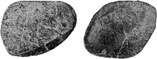
In fishes, the ends of the centra are deeply excavated and connected via a small opening, the passage for the notochord.

In reptiles, this type of centrum is present in embryos, and in adult forms of some species; in most species including dinosaurs, centra are more ossified with the notochordal opening closed, improving resistance against compressional forces.

In long necks and tails, this stabilization works best when the convex part is pointing away from the body.

In sauropods, vertebrae in front of the sacrum are therefore typically opisthocoelous, while those of the tail are procoelous.

Comparison between the air sacs of Majungasaurus and a bird
Skull diagram of the basal sauropodomorph Massospondylus , showing the main skull openings ( fenestra ), including the external naris , the antorbital fenestra , the orbit , the infratemporal fenestra (here: lateral temporal fenestra), the supratemporal fenestra , and the mandibular fenestra .
Bones of the forearm and manus (A), pes (B), and carpus (C) of Heterodontosaurus . ol – olecranon process , ul – ulna , ra – radius , pi – pisiform , ule – ulnare , rae – radiale , mc – metacarpal , ph – phalanx , un – ungual , mt – metatarsal .
Cross-section of the tail of Carnotaurus , showing the tail musculature.
Classification of centrum morphologies based on their articular ends in dinosaurs.
Illustration of cervicals 15-2 of Diplodocus
Chevron of Edmontosaurus in front view.
Skull of Utahceratops . The pronounced coronoid process is labeled "cp".
Lower jaw of Edmontosaurus showing the dental battery
Appendicular skeleton of the basal sauropod Spinophorosaurus . A–C, Tail club osteoderm . D, left coracoid showing the coracoid foramen . E, left scapula showing the acromion . F–G, right humerus showing the deltopectoral crest . H, left femur showing the greater trochanter , the lesser trochanter , and the fourth trochanter . I, left pubis showing the obturator foramen . J, left ischium . K–L, left tibia showing the cnemial crest . M, left fibula .
Gastralia of Poekilopleuron
Humeri of Diamantinasaurus , shown at multiple angles
Left ilium of the basal sauropodomorph Sarahsaurus in lateral view. b – iliac blade , poap – postacetabular process , brv – brevis fossa , isp – ischiadic peduncle , atm – margin of acetabulum , pup – pubic peduncle , sac – supraacetabular ridge , pap – preacetabular process .
Diagram of the forelimb of Allosaurus showing the principal segments of the limb
Skeletal reconstruction of the pachycephalosaur Homalocephale , showing a caudal basket of myorhabdoi of the tail
Osteoderm of Ankylosaurus
Braincase of the sauropod Spinophorosaurus in rear view, showing the postparietal foramen (PPF) and the posttemporal fenestra (PTF)
Ribs and a dorsal vertebra of Ankylosaurus
Variation in neural spine sails across Dinosauria, clockwise from bottom left: Acrocanthosaurus , Amargasaurus , Spinosaurus , Limaysaurus , Ichthyovenator , and Ouranosaurus
Annotated skull diagram of Dromaeosaurus in lateral view showing bones and skull openings.
Diagram of three taxa displaying subnarial gaps.
Thagomizer of Stegosaurus