Keratin
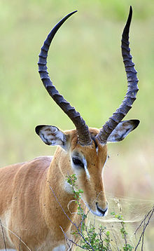
It is the key structural material making up scales, hair, nails, feathers, horns, claws, hooves, and the outer layer of skin in vertebrates.
They form the hair (including wool), the outer layer of skin, horns, nails, claws and hooves of mammals, and the slime threads of hagfish.
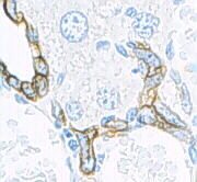
These antibodies are used as fluorescent markers to distinguish subsets of mouse thymic epithelial cells in genetic studies of the thymus.
[17] By analysis of the primary structures of these keratins and other intermediate filament proteins, Hanukoglu and Fuchs suggested a model in which keratins and intermediate filament proteins contain a central ~310 residue domain with four segments in α-helical conformation that are separated by three short linker segments predicted to be in beta-turn conformation.
[20] The major force that keeps the coiled-coil structure is hydrophobic interactions between apolar residues along the keratin's helical segments.
[21] Limited interior space is the reason why the triple helix of the (unrelated) structural protein collagen, found in skin, cartilage and bone, likewise has a high percentage of glycine.
[22] A preponderance of amino acids with small, nonreactive side groups is characteristic of structural proteins, for which H-bonded close packing is more important than chemical specificity.
In addition to intra- and intermolecular hydrogen bonds, the distinguishing feature of keratins is the presence of large amounts of the sulfur-containing amino acid cysteine, required for the disulfide bridges that confer additional strength and rigidity by permanent, thermally stable crosslinking[23]—in much the same way that non-protein sulfur bridges stabilize vulcanized rubber.
Extensive disulfide bonding contributes to the insolubility of keratins, except in a small number of solvents such as dissociating or reducing agents.
Thiolated polymers (thiomers) can form disulfide bridges with cysteine substructures of keratins getting covalently attached to these proteins.
[25] Thiomers therefore exhibit high binding properties to keratins found in hair,[26] on skin[27][28] and on the surface of many cell types.
Eventually the nucleus and cytoplasmic organelles disappear, metabolism ceases and cells undergo a programmed death as they become fully keratinized.
Rubbing and pressure cause thickening of the outer, cornified layer of the epidermis and form protective calluses, which are useful for athletes and on the fingertips of musicians who play stringed instruments.
Silk found in insect pupae, and in spider webs and egg casings, also has twisted β-pleated sheets incorporated into fibers wound into larger supermolecular aggregates.
The structure of the spinnerets on spiders' tails, and the contributions of their interior glands, provide remarkable control of fast extrusion.
Silk from the hornet cocoon contains doublets about 10 μm across, with cores and coating, and may be arranged in up to 10 layers, also in plaques of variable shape.
Cats regularly ingest hair as part of their grooming behavior, leading to the gradual formation of hairballs that may be expelled orally or excreted.