In molecular biology, the term macromolecular assembly (MA) refers to massive chemical structures such as viruses and non-biologic nanoparticles, cellular organelles and membranes and ribosomes, etc.
Macromolecules are found in living and nonliving things, and are composed of many hundreds or thousands of atoms held together by covalent bonds; they are often characterized by repeating units (i.e., they are polymers).
[6] Examples: The biomacromolecular complexes are studied structurally by X-ray crystallography, NMR spectroscopy of proteins, cryo-electron microscopy and successive single particle analysis, and electron tomography.
In addition, they play fundamental roles in all basic life processes (protein translation, cell division, vesicle trafficking, intra- and inter-cellular exchange of material between compartments, etc.).
[citation needed] Virus structures were among the first studied MAs; other biologic examples include ribosomes (partial image above), proteasomes, and translation complexes (with protein and nucleic acid components), procaryotic and eukaryotic transcription complexes, and nuclear and other biological pores that allow material passage between cells and cellular compartments.
The crystallization and structure solution for the ribosome, MW ~ 2.5 MDa, an example of part of the protein synthetic 'machinery' of living cells, was object of the 2009 Nobel Prize in Chemistry awarded to Venkatraman Ramakrishnan, Thomas A. Steitz, and Ada E.
Structure of nucleoprotein MA: The 50S ribosomal subunit from
H. marismortui
X-ray crystallographic
model of 29 of the 33 native components, from the laboratory of
Thomas Steitz
. Of the 31 component proteins, 27 are shown (blue), along with its 2 RNA strands (orange/yellow).
[
1
]
Scale: assembly is approx. 24 nm across.
[
2
]
A eukaryotic
ribosome
, which catalytically
translate
the information content contained in
mRNA
molecules into proteins. The animation presents the elongation and membrane targeting stages of
eukaryotic translation
, showing the mRNA as a black arc, the
ribosome
subunits in green and yellow, tRNAs in dark blue, proteins such as
elongation
and other factors involved in light blue, the growing polypeptide chain as a black thread growing vertically from the curve of the mRNA. At end of the animation, the polypeptide produced is extruded through a light blue SecY pore
[
3
]
into the gray interior of the
ER
.
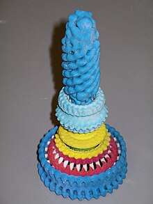
3D printed
model of the structure of a
bacterial
flagellum
"motor" and partial rod structure of a
Salmonella
species. Bottom to top: dark blue, repeating FliM and FliN, motor/switch proteins; red, FliG motor/switch proteins; yellow, FliF transmembrane coupling proteins; light blue, L and P ring proteins; and (at top), dark blue, the cap, hook-filament junction, hook, and rod proteins.
[
4
]
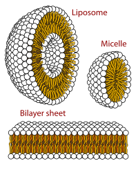
Cross-sections of phospholipid (PLs) relevant to
biomembrane
MAs. Yellow-orange indicates
hydrophobic
lipid tails; black and white spheres represent PL polar regions (
v.i.
). Bilayer/liposome dimensions (obscured in graphic): hydrophobic and polar regions, each ~30 Å (3.0 nm) "thick"—the polar from ~15 Å (1.5 nm)
on each side
.
[
11
]
[
12
]
[
13
]
[
non-primary source needed
]
[
14
]
A graphical representation of the structure of a viral MA,
cowpea mosaic virus
, with 30 copies of each of its coat proteins, the small coat protein (S, yellow) and the large coat protein (L, green), which, along with 2 molecules of
positive-sense
RNA
(RNA-1 and RNA-2, not visible) constitute the virion. The assembly is highly
symmetric
, and is ~280 Å (28 nm) across at its widest point.
[
verification needed
]
[
citation needed
]