Opsin
Animal opsins are G-protein-coupled receptors and a group of proteins made light-sensitive via a chromophore, typically retinal.
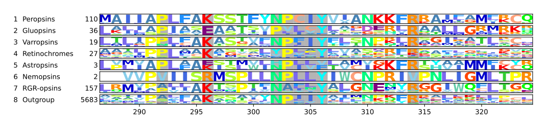
Five classical groups of opsins are involved in vision, mediating the conversion of a photon of light into an electrochemical signal, the first step in the visual transduction cascade.
Another opsin found in the mammalian retina, melanopsin, is involved in circadian rhythms and pupillary reflex but not in vision.
Opsins are G-protein-coupled receptors (GPCRs),[1][2] which are chemoreceptors and have seven transmembrane domains forming a binding pocket for a ligand.
[3][4] The ligand for opsins is the vitamin A-based chromophore 11-cis-retinal,[5][6][7][8][9] which is covalently bound to a lysine residue[10] in the seventh transmembrane domain[11][12][13] through a Schiff-base.
For instance in the seventh transmembrane domain, the proline in the highly conserved NPxxY7.53 motif is Pro7.50, the asparagine before is then Asp7.49, and the tyrosine three residues after is then Tyr7.53.
[33][34] In particular, the Drosophila rhabdomeric opsins (rhabopsins, r-opsins) Rh1, Rh4, and Rh7 function not only as photoreceptors, but also as chemoreceptors for aristolochic acid.
Therefore, RGR-opsins and retinochromes are thought to neither signal nor activate a phototransduction cascade but to work as photoisomerases to produce 11-cis-retinal for other opsins.
Certain amino acid residues, termed spectral tuning sites, have a strong effect on λmax values.
Using site-directed mutagenesis, it is possible to selectively mutate these residues and investigate the resulting changes in light absorption properties of the opsin.
They are not mutually exclusive, but, for practical reasons, it is easier to investigate spectral tuning sites that do not affect opsin functionality.
Frogs are distinct among vertebrates because they lack the RH2 opsin, typically used for detecting middle wavelengths of light in other species.
This loss likely reflects their evolutionary focus on low-light vision, with RH1, a rod-specific opsin, taking the lead in supporting nocturnal and crepuscular (dawn and dusk) activity.
The SWS2 opsin, for instance, is tuned to detect blue and green light, which is especially useful in aquatic environments or shaded areas.
This tuning is enhanced by specific mutations which increases sensitivity to low-light conditions and stabilizes the protein for better performance in dim environments.
This change aligns with their reliance on longer wavelengths, like red and yellow, for tasks such as mate selection and predator deterrence, often linked to their vibrant aposematic (warning) coloration.
[69] They convert light signals to nerve impulses via cyclic nucleotide gated ion channels, which work by increasing the charge differential across the cell membrane (i.e.
They are further subdivided into: These pineal opsins, found in the Actinopterygii (ray-finned fish) apparently arose as a result of gene duplication from Rh1 (rhodopsin).
[83][84] The panopsins are found in many tissues (skin,[51] brain,[53][85] testes,[53] heart, liver,[85] kidney, skeletal muscle, lung, pancreas and retina[85]).
[53] The first invertebrate panopsin was found in the ciliary photoreceptor cells of the annelid Platynereis dumerilii and is called c(iliary)-opsin.
The color unlike brightness stays almost constant independent of time of day or the weather, for instance if it is cloudy.
[96] Cnidaria, which include jellyfish, corals, and sea anemones, are the most basal animals to possess complex eyes.
[21][28] However, earlier studies have found that some cnidarian opsins belong to the cilopsins, rhabopsins, and the tetraopsins of the bilaterians.
[101] Melanopsin (OPN4) is involved in circadian rhythms, the pupillary reflex, and color correction in high-brightness situations.
), type of chromophore (retinal, flavine, bilin), molecular structure (tertiary, quaternary), signal output (phosphorylation, reduction, oxidation), etc.
Both types are called opsins, because at one time it was thought that they were related: Both are seven-transmembrane receptors and bind covalently retinal as chromophore, which turns them into photoreceptors sensing light.
[116] In fact, the sequence identity between animal and mirobial opsins is no greater than could be accounted for by random chance.
The retinal chromophore is found solely in the opsin branch of this large gene family, meaning its occurrence elsewhere represents convergent evolution, not homology.
[122] Several microbal opsins, such as proteo- and bacteriorhodopsin, are used by various bacterial groups to harvest energy from light to carry out metabolic processes using a non-chlorophyll-based pathway.
Beside that, halorhodopsins of Halobacteria and channelrhodopsins of some algae, e.g. Volvox, serve them as light-gated ion channels, amongst others also for phototactic purposes.