Rutherford backscattering spectrometry
Sometimes referred to as high-energy ion scattering (HEIS) spectrometry, RBS is used to determine the structure and composition of materials by measuring the backscattering of a beam of high energy ions (typically protons or alpha particles) impinging on a sample.
Rutherford supervised a series of experiments carried out by Hans Geiger and Ernest Marsden between 1909 and 1914 studying the scattering of alpha particles through metal foils.
Instead, when Marsden positioned the detector on the same side of the foil as the alpha particle source, he immediately detected a noticeable backscattered signal.
"[1] Rutherford interpreted the result of the Geiger–Marsden experiment as an indication of a Coulomb collision with a single massive positive particle.
This model was eventually superseded by the Bohr atom, incorporating some early results from quantum mechanics.
This may result in nuclear reactions in certain cases, but frequently the interaction remains elastic, although the scattering cross-sections may fluctuate wildly as a function of energy and no longer be calculable analytically.
There has recently been great progress in determining EBS scattering cross-sections, by solving Schrödinger's equation for each interaction[citation needed].
However, for the EBS analysis of matrices containing light elements, the utilization of experimentally measured[2][3] scattering cross-section data is also considered to be a very credible option.
This is the basis of the Elastic Recoil Detection (ERD, with synonyms ERDA, FRS, HFS) technique.
The kinematical factor must remain real, and this limits the permitted scattering angle in the laboratory frame of reference.
If the scattering cross-section is zero it implies that the projectile never comes close to the target, but in this case it also never penetrates the electron cloud surrounding the nucleus either.
The pure Coulomb formula for the scattering cross-section shown above must be corrected for this screening effect, which becomes more important as the energy of the projectile decreases (or, equivalently, its mass increases).
The amount by which the ion energy is lowered after passing through a given distance is referred to as the stopping power of the material and is dependent on the electron distribution.
It is generally given in thin film units, that is eV /(atom/cm2) since it is measured experimentally on thin films whose thickness is always measured absolutely as mass per unit area, avoiding the problem of determining the density of the material which may vary as a function of thickness.
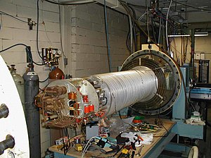
This arrangement is simple and convenient, but it can be difficult to achieve energies of much more than 1 MeV due to the difficulty of applying very high voltages to the system.
The ions thus start out being attracted to the terminal, pass through and become positive, and are repelled until they exit the tube at ground.
Instead it creates a gradual energy loss dependent on the electron density and the distance traversed in the sample.
The result is that instead of the sharp backscattered peaks one would expect on an N(E) plot, with the width determined by energy and angular resolution, the peaks observed trail off gradually towards lower energy as the ions pass through the depth occupied by that element.
Elements which only appear at some depth inside the sample will also have their peak positions shifted by some amount which represents the distance an ion had to traverse to reach those nuclei.
To fully understand the interaction of an incident beam of nuclei with a crystalline structure, it is necessary to comprehend two more key concepts: blocking and channeling.
This occurs because the repulsive potential of the target atom bends close ion trajectories away from their original path, and is referred to as blocking.
This can result in a drastic reduction of the observed backscattered signal when the incident beam is oriented along one of the symmetry directions, allowing determination of a sample's regular crystal structure.
The tolerance for the deviation of the ion beam angle of incidence relative to the symmetry direction depends on the blocking radius, making the allowable deviation angle proportional to While the intensity of an RBS peak is observed to decrease across most of its width when the beam is channeled, a narrow peak at the high-energy end of larger peak will often be observed, representing surface scattering from the first layer of atoms.
[10] If atoms within the target are displaced from their crystalline lattice site, this will result in a higher backscattering yield in relation to a perfect crystal.
By comparing the spectrum from a sample being analyzed to that from a perfect crystal, and that obtained at a random (non-channeling) orientation (representative of a spectrum from an amorphous sample), it is possible to determine the extent of crystalline damage in terms of a fraction of displaced atoms.
A well-known example of this is the RBS analysis of the premelting of lead surfaces by Frenken, Maree and van der Veen.
This increase in the disorder of the surface, making deeper atoms visible to the incident beam, was interpreted as pre-melting of the surface, and computer simulations of the RBS process produced similar results when compared with theoretical pre-melting predictions.

Right: Observed results: a small portion of the particles were deflected, indicating a small, concentrated positive charge.