Sensory nervous system
Commonly recognized sensory systems are those for vision, hearing, touch, taste, smell, balance and visceral sensation.
The receptors which react to the stimulus and initiate the process of sensation are commonly characterized in four distinct categories: chemoreceptors, photoreceptors, mechanoreceptors, and thermoreceptors.
The two primary types of chemoreceptors are: Photoreceptors are neuron cells and are specialized units that play the main role in initiating vision function.
Different types of photoreceptors are able to respond to the varying light wavelengths in relation to color, and transduce them into electrical signals.
[6] Photoreceptors are capable of phototransduction, a process which converts light (electromagnetic radiation) into, among other types of energy, a membrane potential.
As its name suggests, CC works to connect the OS and the IS regions together for the purpose of essential protein trafficking.
In humans, rods outnumber cones by approximately 20:1, while in nocturnal animals, such as the tawny owl, the ratio is closer to 1000:1.
While the mechanisms through which these receptors operate is unclear, recent discoveries have shown that mammals have at least two distinct types of thermoreceptors:[14] TRPV1 is a heat-activated channel that acts as a small heat detecting thermometer in the membrane which begins the polarization of the neural fiber when exposed to changes in temperature.
Both cold and hot receptors are segregated by distinct subpopulations of sensory nerve fibers, which shows us that the information coming into the spinal cord is originally separate.
[15] Nociceptors respond to potentially damaging stimuli by sending signals to the spinal cord and brain.
All stimuli received by the receptors listed above are transduced to an action potential, which is carried along one or more afferent neurons towards a specific area of the brain.
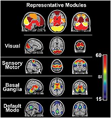
'[20] Increases in task-negative activity are observed in the ventral attention network, after abrupt changes in sensory stimuli,[21] at the onset and offset of task blocks,[22] and at the end of a completed trial.[23][relevant?]
Located in the temporal lobe, the auditory cortex is the primary receptive area for sound information.
Unique to the olfactory and gustatory systems, at least in mammals, is the implementation of both peripheral and central mechanisms of action.
In contrast, the term flavor refers to the experience generated through integration of taste with smell and tactile information.
[clarification needed] Peripheral taste receptors, located on the tongue, soft palate, pharynx, and esophagus, transmit the received signal to primary sensory axons, where the signal is projected to the nucleus of the solitary tract in the medulla, or the gustatory nucleus of the solitary tract complex.
Scent, in contrast, is not combined with taste to create flavor until higher cortical processing regions, such as the insula and orbitofrontal cortex.
[citation needed] It is not always well-defined for nonlinear, nonpassive sensory organs, since they can't function without input energy.
[citation needed] Some sensory systems can have multiple quiescent states depending on its history, like flip-flops, and magnetic material with hysteresis.
[27] Quiescent state is less well-defined when the sensory organ can be controlled by other systems, like a dog's ears that turn towards the front or the sides as the brain commands.