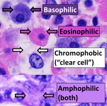
Basophilic
It describes the appearance of cells, tissues and cellular structures as seen through the microscope after a histological section has been stained with a basic dye.
Basic dyes are cationic, i.e. contain positive charges, and thus they stain anionic structures (i.e. structures containing negative charges), such as the phosphate backbone of DNA in the cell nucleus and ribosomes.
[1] "Basophils" are cells that "love" (from greek "-phil") basic dyes, for example haematoxylin, azure and methylene blue.
Specifically, this term refers to: An abnormal increase in basophil granulocytes is therefore also described as basophilia.
These structures contain many positive charges and are thus strongly stained by anionic dyes like eosin.