Molecular graphics
[6] Thermal ellipsoid plots quickly became the de facto standard used in the display of X-ray crystallography data, and are still in wide use today.
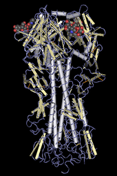
[6] The first practical use of molecular graphics was a simple display of the protein myoglobin using a wireframe representation in 1966 by Cyrus Levinthal and Robert Langridge working at Project MAC.
[7] Among the milestones in high-performance molecular graphics was the work of Nelson Max in "realistic" rendering of macromolecules using reflecting spheres.
Stereoscopic viewing glasses were designed using lead lanthanum zirconate titanate (PLZT) ceramics as electronically-controlled shutter elements.
The first two protein structures solved by molecular graphics without the aid of the Richards' Box were built with Stan Swanson's program FIT on the Vector General graphics display in the laboratory of Edgar Meyer at Texas A&M University: First Marge Legg in Al Cotton's lab at A&M solved a second, higher-resolution structure of staph.
In the space-filling model, atoms are drawn as solid spheres to suggest the space they occupy, in proportion to their van der Waals radii.