Blood smear
The monolayer is found in the "feathered edge" created by the spreader slide as it draws the blood forward.
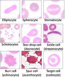
[3] Microscopic examination of the shape, size, and coloration of red blood cells is useful for determining the cause of anemia.
[5][6] Blood smear examination is the preferred diagnostic method for certain parasitic infections, such as malaria and babesiosis.
[8] The preferred and most reliable diagnosis of malaria is microscopic examination of blood smears, because each of the four major parasite species has distinguishing characteristics.
As blood cools to room temperature, male gametocytes will divide and release microgametes: these are long sinuous filamentous structures that can be mistaken for organisms such as Borrelia.
If the blood is kept at warmer temperatures, schizonts will rupture and merozoites invading erythrocytes will mistakenly give the appearance of the accolé form of P. falciparum.
If P. vivax or P. ovale is left for several hours in EDTA, the buildup of acid in the sample will cause the parasitised erythrocytes to shrink and the parasite will roll up, simulating the appearance of P. malariae.