Action potential

[b] When the channels open, they allow an inward flow of sodium ions, which changes the electrochemical gradient, which in turn produces a further rise in the membrane potential towards zero.

Action potentials are driven by channel proteins whose configuration switches between closed and open states as a function of the voltage difference between the interior and exterior of the cell.

In the Hodgkin–Huxley membrane capacitance model, the speed of transmission of an action potential was undefined and it was assumed that adjacent areas became depolarized due to released ion interference with neighbouring channels.

[26][27][28][29] In sensory neurons, an external signal such as pressure, temperature, light, or sound is coupled with the opening and closing of ion channels, which in turn alter the ionic permeabilities of the membrane and its voltage.

For illustration, in the human ear, hair cells convert the incoming sound into the opening and closing of mechanically gated ion channels, which may cause neurotransmitter molecules to be released.

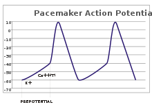
[g] Although such pacemaker potentials have a natural rhythm, it can be adjusted by external stimuli; for instance, heart rate can be altered by pharmaceuticals as well as signals from the sympathetic and parasympathetic nerves.

[7][10] A complicating factor is that a single ion channel may have multiple internal "gates" that respond to changes in Vm in opposite ways, or at different rates.

The voltages and currents of the action potential in all of its phases were modeled accurately by Alan Lloyd Hodgkin and Andrew Huxley in 1952,[i] for which they were awarded the Nobel Prize in Physiology or Medicine in 1963.

[lower-Greek 2] However, their model considers only two types of voltage-sensitive ion channels, and makes several assumptions about them, e.g., that their internal gates open and close independently of one another.

[43][44][45] At longer times, after some but not all of the ion channels have recovered, the axon can be stimulated to produce another action potential, but with a higher threshold, requiring a much stronger depolarization, e.g., to −30 mV.

[39] Combined, these changes in sodium and potassium permeability cause Vm to drop quickly, repolarizing the membrane and producing the "falling phase" of the action potential.

When closing after an action potential, sodium channels enter an "inactivated" state, in which they cannot be made to open regardless of the membrane potential—this gives rise to the absolute refractory period.

At the molecular level, this absolute refractory period corresponds to the time required for the voltage-activated sodium channels to recover from inactivation, i.e., to return to their closed state.

In order to enable fast and efficient transduction of electrical signals in the nervous system, certain neuronal axons are covered with myelin sheaths.

For axons larger than a minimum diameter (roughly 1 micrometre), myelination increases the conduction velocity of an action potential, typically tenfold.

[69] It is likely that the familiar signaling function of action potentials in some vascular plants (e.g. Mimosa pudica) arose independently from that in metazoan excitable cells.

[ap] Sponges seem to be the main phylum of multicellular eukaryotes that does not transmit action potentials, although some studies have suggested that these organisms have a form of electrical signaling, too.

The initial work, prior to 1955, was carried out primarily by Alan Lloyd Hodgkin and Andrew Fielding Huxley, who were, along John Carew Eccles, awarded the 1963 Nobel Prize in Physiology or Medicine for their contribution to the description of the ionic basis of nerve conduction.

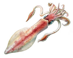
[ar] These axons are so large in diameter (roughly 1 mm, or 100-fold larger than a typical neuron) that they can be seen with the naked eye, making them easy to extract and manipulate.

[av][aw] Refinements of this method are able to produce electrode tips that are as fine as 100 Å (10 nm), which also confers high input impedance.

[76] Action potentials may also be recorded with small metal electrodes placed just next to a neuron, with neurochips containing EOSFETs, or optically with dyes that are sensitive to Ca2+ or to voltage.

Optical imaging technologies have been developed in recent years to measure action potentials, either via simultaneous multisite recordings or with ultra-spatial resolution.

[bb] In the 19th century scientists studied the propagation of electrical signals in whole nerves (i.e., bundles of neurons) and demonstrated that nervous tissue was made up of cells, instead of an interconnected network of tubes (a reticulum).

Matteucci's work inspired the German physiologist, Emil du Bois-Reymond, who discovered in 1843 that stimulating these muscle and nerve preparations produced a notable diminution in their resting currents, making him the first researcher to identify the electrical nature of the action potential.

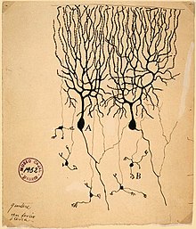
To establish that nervous tissue is made up of discrete cells, the Spanish physician Santiago Ramón y Cajal and his students used a stain developed by Camillo Golgi to reveal the myriad shapes of neurons, which they rendered painstakingly.

[bc][80] Bernstein's hypothesis was confirmed by Ken Cole and Howard Curtis, who showed that membrane conductance increases during an action potential.

[bd] In 1907, Louis Lapicque suggested that the action potential was generated as a threshold was crossed,[be] what would be later shown as a product of the dynamical systems of ionic conductances.

[i] Hodgkin and Huxley correlated the properties of their mathematical model with discrete ion channels that could exist in several different states, including "open", "closed", and "inactivated".

Their hypotheses were confirmed in the mid-1970s and 1980s by Erwin Neher and Bert Sakmann, who developed the technique of patch clamping to examine the conductance states of individual ion channels.

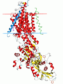
[h] The sodium–potassium pump was identified in 1957[bl][lower-Greek 6] and its properties gradually elucidated,[bm][bn][bo] culminating in the determination of its atomic-resolution structure by X-ray crystallography.

As an action potential (nerve impulse) travels down an axon there is a change in electric polarity across the membrane of the axon. In response to a signal from another neuron , sodium- (Na + ) and potassium- (K + )–gated ion channels open and close as the membrane reaches its threshold potential . Na + channels open at the beginning of the action potential, and Na + moves into the axon, causing depolarization . Repolarization occurs when K + channels open and K + moves out of the axon, creating a change in electric polarity between the outside of the cell and the inside. The impulse travels down the axon in one direction only, to the axon terminal where it signals other neurons.
Shape of a typical action potential. The membrane potential remains near a baseline level until at some point in time, it abruptly spikes upward and then rapidly falls.
Approximate plot of a typical action potential shows its various phases as the action potential passes a point on a cell membrane . The membrane potential starts out at approximately −70 mV at time zero. A stimulus is applied at time = 1 ms, which raises the membrane potential above −55 mV (the threshold potential). After the stimulus is applied, the membrane potential rapidly rises to a peak potential of +40 mV at time = 2 ms. Just as quickly, the potential then drops and overshoots to −90 mV at time = 3 ms, and finally the resting potential of −70 mV is reestablished at time = 5 ms.
Action potential propagation along an axon
Ion movement during an action potential.
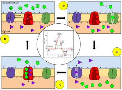
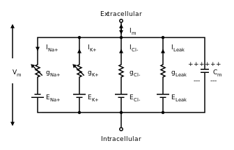
Key: a) Sodium (Na + ) ion. b) Potassium (K + ) ion. c) Sodium channel. d) Potassium channel. e) Sodium-potassium pump.
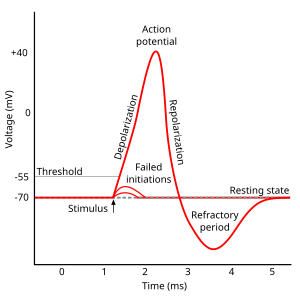
In the stages of an action potential, the permeability of the membrane of the neuron changes. At the resting state (1), sodium and potassium ions have limited ability to pass through the membrane, and the neuron has a net negative charge inside. Once the action potential is triggered, the depolarization (2) of the neuron activates sodium channels, allowing sodium ions to pass through the cell membrane into the cell, resulting in a net positive charge in the neuron relative to the extracellular fluid. After the action potential peak is reached, the neuron begins repolarization (3), where the sodium channels close and potassium channels open, allowing potassium ions to cross the membrane into the extracellular fluid, returning the membrane potential to a negative value. Finally, there is a refractory period (4), during which the voltage-dependent ion channels are inactivated while the Na + and K + ions return to their resting state distributions across the membrane (1), and the neuron is ready to repeat the process for the next action potential.
The pre- and post-synaptic axons are separated by a short distance known as the synaptic cleft. Neurotransmitter released by pre-synaptic axons diffuse through the synaptic clef to bind to and open ion channels in post-synaptic axons.
When an action potential arrives at the end of the pre-synaptic axon (top), it causes the release of neurotransmitter molecules that open ion channels in the post-synaptic neuron (bottom). The combined excitatory and inhibitory postsynaptic potentials of such inputs can begin a new action potential in the post-synaptic neuron.
A plot of action potential (mV) vs time. The membrane potential is initially −60 mV, rise relatively slowly to the threshold potential of −40 mV, and then quickly spikes at a potential of +10 mV, after which it rapidly returns to the starting −60 mV potential. The cycle is then repeated.
In pacemaker potentials , the cell spontaneously depolarizes (straight line with upward slope) until it fires an action potential.
Axons of neurons are wrapped by several myelin sheaths, which shield the axon from extracellular fluid. There are short gaps between the myelin sheaths known as nodes of Ranvier where the axon is directly exposed to the surrounding extracellular fluid.
In saltatory conduction , an action potential at one node of Ranvier causes inwards currents that depolarize the membrane at the next node, provoking a new action potential there; the action potential appears to "hop" from node to node.
A log-log plot of conduction velocity (m/s) vs axon diameter (μm).
Comparison of the conduction velocities of myelinated and unmyelinated axons in the cat . [ 55 ] The conduction velocity v of myelinated neurons varies roughly linearly with axon diameter d (that is, v d ), [ p ] whereas the speed of unmyelinated neurons varies roughly as the square root ( v d ). [ u ] The red and blue curves are fits of experimental data, whereas the dotted lines are their theoretical extrapolations.
A diagram showing the resistance and capacitance across the cell membrane of an axon. The cell membrane is divided into adjacent regions, each having its own resistance and capacitance between the cytosol and extracellular fluid across the membrane. Each of these regions is in turn connected by an intracellular circuit with a resistance.
Cable theory's simplified view of a neuronal fiber. The connected RC circuits correspond to adjacent segments of a passive neurite . The extracellular resistances r e (the counterparts of the intracellular resistances r i ) are not shown, since they are usually negligibly small; the extracellular medium may be assumed to have the same voltage everywhere.
Electrical synapases are composed of protein complexes that are imbedded in both membranes of adjacent neurons and thereby provide a direct channel for ions to flow from the cytoplasm of one cell into an adjacent cell.
Electrical synapses between excitable cells allow ions to pass directly from one cell to another, and are much faster than chemical synapses .
Plot of membrane potential versus time. The initial resting phase (region 4) is negative and constant flowed by sharp rise (0) to a peak (1). The plateau phase (2) is slightly below the peak. The plateau phase is followed by a fairly rapid return (3) back to the resting potential (4).
Phases of a cardiac action potential. The sharp rise in voltage ("0") corresponds to the influx of sodium ions, whereas the two decays ("1" and "3", respectively) correspond to the sodium-channel inactivation and the repolarizing eflux of potassium ions. The characteristic plateau ("2") results from the opening of voltage-sensitive calcium channels.
Illustration of the longfin inshore squid.
Giant axons of the longfin inshore squid ( Doryteuthis pealeii ) were crucial for scientists to understand the action potential. [ 73 ]
Plot of membrane potential versus time. The channel is primarily in a high conductance state punctuated by random and relatively brief transitions to a low conductance states
As revealed by a patch clamp electrode, an ion channel has two states: open (high conductance) and closed (low conductance).
Photograph of a pufferfish.
Tetrodotoxin is a lethal toxin found in pufferfish that inhibits the voltage-sensitive sodium channel , halting action potentials.
Hand drawn figure of two Purkinje cells side by side with dendrites projecting upwards that look like tree branches and a few axons projected downwards that connect to a few granule cells at the bottom of the drawing.
Image of two Purkinje cells (labeled as A ) drawn by Santiago Ramón y Cajal in 1899. Large trees of dendrites feed into the soma , from which a single axon emerges and moves generally downwards with a few branch points. The smaller cells labeled B are granule cells .
Cartoon diagram of the sodium–potassium pump drawn vertically imbedded in a schematic diagram of a lipid bilayer represented by two parallel horizontal lines. The portion of the protein that is imbedded in the lipid bilayer is composed largely of anti-parallel beta sheets. There is also a large intracellular domain of the protein with a mixed alpha-helix/beta-sheet structure.
Ribbon diagram of the sodium–potassium pump in its E2-Pi state. The estimated boundaries of the lipid bilayer are shown as blue (intracellular) and red (extracellular) planes.
Circuit diagram depicting five parallel circuits that are interconnected at the top to the extracellular solution and at the bottom to the intracellular solution.
Equivalent electrical circuit for the Hodgkin–Huxley model of the action potential. I m and V m represent the current through, and the voltage across, a small patch of membrane, respectively. The C m represents the capacitance of the membrane patch, whereas the four g 's represent the conductances of four types of ions. The two conductances on the left, for potassium (K) and sodium (Na), are shown with arrows to indicate that they can vary with the applied voltage, corresponding to the voltage-sensitive ion channels . The two conductances on the right help determine the resting membrane potential .