H&E stain
[8] The results from H&E staining are not overly dependent on the chemical used to fix the tissue or slight inconsistencies in laboratory protocol,[11] and these factors contribute to its routine use in histology.
[7] After tissues have been collected (often as biopsies) and fixed, they are typically dehydrated and embedded in melted paraffin wax, the resulting block is mounted on a microtome and cut into thin slices.
[6][8][7] Hematoxylin principally colors the nuclei of cells blue or dark-purple,[6][15][14] along with a few other tissues, such as keratohyalin granules and calcified material.
[8][13] Hematoxylin, when combined with a mordant (most commonly aluminum alum) is often considered to "resemble"[10] a basic, positively charged, or cationic stain.
There is evidence to indicate that co-ordinate bonds, similar to those that hold aluminium and hematein together, bind the hemalum complex to DNA and to carboxy groups of proteins in the nuclear chromatin.
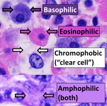
[citation needed] The structures do not have to be acidic or basic to be called basophilic and eosinophilic; the terminology is based on the affinity of cellular components for the dyes.
Hydrophobic structures also tend to remain clear; these are usually rich in fats, e.g. adipocytes, myelin around neuron axons, and Golgi apparatus membranes.